Telomere length is a very precise tool to determine the physiological age of our cells and organs.
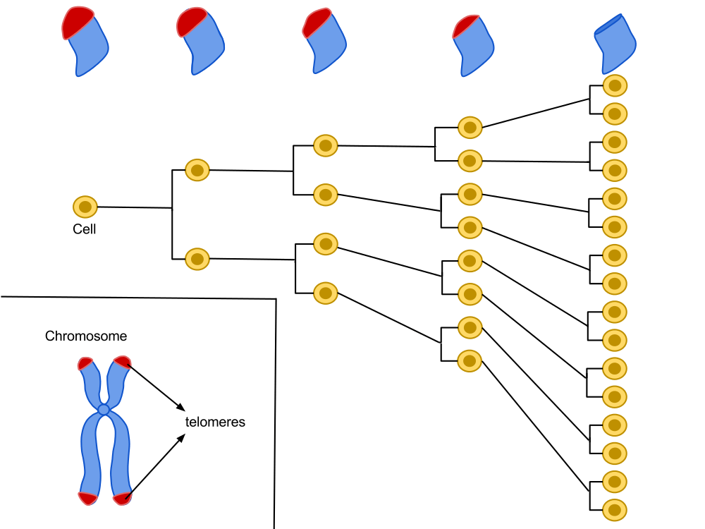
Located at the very end of our chromosomes, telomeres are repetitive sequences of the nucleotide suite TTAGGG. A cell is programed for a given number of divisions. With each division, the ends of the chromosomes become a little shorter, until they reach a critical size. Through numerous reactions, the cell will then stop growing and become senescent.
Lifestyle, diet, stress, environment as well as age can accelerate telomere shortening. Measuring their length can then give us some precious indications on how old our cells really are, as well as what makes them age. For all those reasons, telomere length is a great metrology tool when it comes to measuring the level of aging of an organism, it can influence people into taking control of their health with preventive measures.
Hayflick’s work showed that each cell line has a capital of cellular divisions. This capital is proportional to the longevity of a species and can vary between two individuals of the same species. With each cycle of cell division, chromosome ends (telomeres) lose a fragment of DNA. After several divisions, telomere function, which helps maintain the stability of chromosomes in the DNA, is altered, which could be the basis of our biological clock.
The sample: Telomere length can be measured on cell or tissue samples. However, leucocytes (white blood cells) are the preferred sample for telomere measurement, since they are blood cells and allow for a non-invasive sample collection.
Southern blots of Terminal Restriction Fragments (TRFs)
The most archaic but also the most widely used technique was developed in 1990. It estimates the average number of TTAGGG ending repetitions in the chromosomes.
The technique works by in situ hybridization identifying the TTAGGG repetitions at the end of telomeres. Telomeric DNA is digested by restriction enzymes, which makes for many small DNA segments of different sizes. In order to know the size of each telomeric DNA fragment, the Southern Blot technique allows to separate DNA molecules depending on their length.
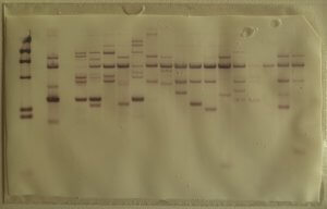
The resulting telomeric fragments are separated by gel electrophoresis. In order to make the rest of the process easier, the fragments are transferred onto a nitrocellulose or nylon membrane, which are solid supports that help denature DNA and hybridizing it with a probe.
This is a crucial step for the hybridization of a radioactive probe (a complementary DNA fragment on the given sequence.) The position of the restriction fragments is revealed by radiography and their size is estimated by comparing the distance they traveled in the gel and the distance traveled by fragments of a known length [1, 2].
Q-Fish
This method also uses in situ hybridization. However, unlike the Souther Blot method, the probe is not radioactive but fluorescent. The drawback with this method is the important quantity of DNA required (up to 20μg) when only a few ng are sufficient for other PCR-based techniques [3, 4].
Flow-FISH
This technique is similar to the Q-FISH technique in many respects, but Flow-Fisch uses a flow cytometer. This rarely used, expensive technique requires a great deal of equipment. However, it remains very interesting: it allows to reduce the purification steps and to simultaneously analyze telomere length in several cell types [7,8].
TAT to measure telomere length
TAT® estimates the individual length of telomeres. The TAT® technology is based on the high flow, in situ fluorescent hybridization allowed by a High Content Screening (HCS) approach of telomere length analysis. It gives a full measure of telomere length through a histogram. It also gives indications on the shorter and longer telomeres, the frequency by length class, the average length as well as the median values [9].
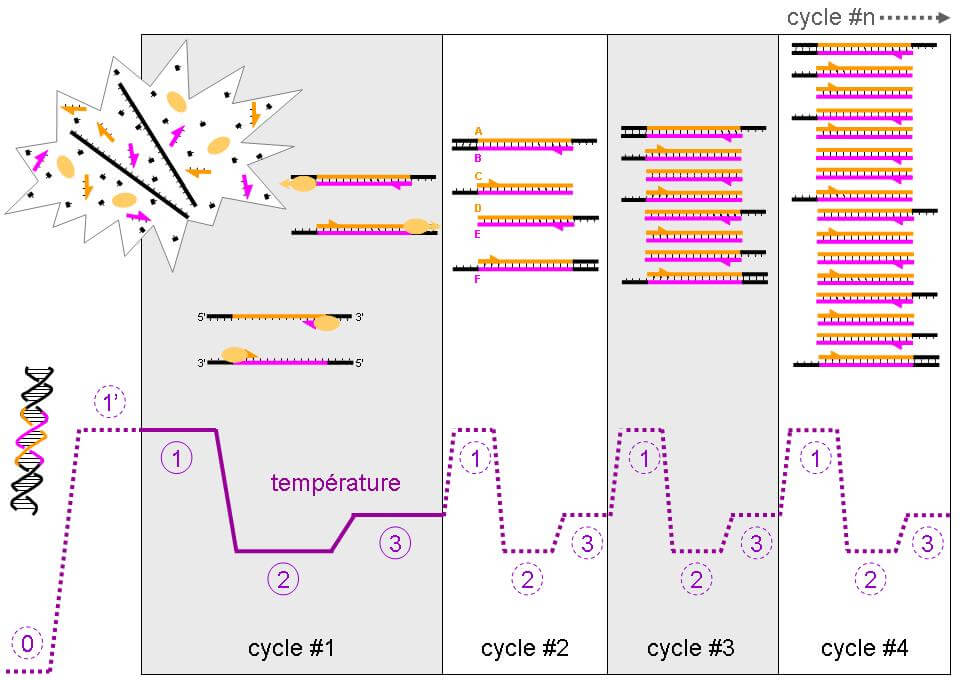
PCR-Q
Faster and easier, the qPCR technique was developed in 2003. It determines by quantitative PCR the amount of copies of the telomere pattern (T) against a single copy gene (S) with a (T/S) ratio. The number of TTACC repetitions, which are specific to telomeric DNA, is given by the kinetic measure of the polymerization reaction. During the amplification process, the amount of amplicons is monitored with a fluorescent marker which helps to obtain the polymerization reaction kinetic, which in turn helps determine the initial quantity of telomeric DNA [2, 5, 6].
STELA (Single TElomere Length Analysis)
Also based on PCR, this technique shows better resolution than the Southern Blot technique. It measures the length of telomeres by linking a primer on a specific chromosome’s telomere {2, 7, 8].
PCR techniques might seem better because easily reproducible. However, like other techniques based on in-situ hybridization, they cannot amplify telomeres over 25 kb. It is possible to observe telomeres longer than 50 kb, but the Southern Blot technique is a better tool for this use. We know that not all organs age at the same pace, and even though leucocytes are representative when it comes to studying aging, these tests mostly analyze cell populations from blood samples, which remains simplistic.
Analyzing telomere length from a tissue biopsy is a possibility, but requires specific and exceptional conditions, such as a medical diagnosis. The estimated age is not based on a follow-up in time, but on a comparison to an average in chronological age.
Where can we measure our telomere length?
Many a laboratory has embraced the market, each commercializing its own version of the telomere length analysis kit. The Life Length company is a leader in the field, and own many partner laboratories offering the analysis of samples through the postal service like Les Laboratoires Réunis, or an on-site analysis like La Clinique Crillon.
This futuristic technology is still in its infancy but makes for great future discoveries in the upcoming years, thanks to both the advances in available methods and the mutliplication of laboratories offering such services. They lower the cost, time and risk margin of these analyses.