• Log in with Facebook Log in with Twitter Log In with Google      Sign In    
  • Create Account
  LongeCity
              Advocacy & Research for Unlimited Lifespans

Photo

De novo mutations in mitochondrial DNA of iPSCs produce immunogenic neoepitopes in mice and humans

stem cell research transplant immunology

  • Please log in to reply
1 reply to this topic

#1 Engadin

  • Guest
  • 198 posts
  • 580
  • Location:Madrid
  • NO

Posted 22 August 2019 - 02:19 PM


 S O U R C E :   n a t u r e (ePDF)


ABSTRACT


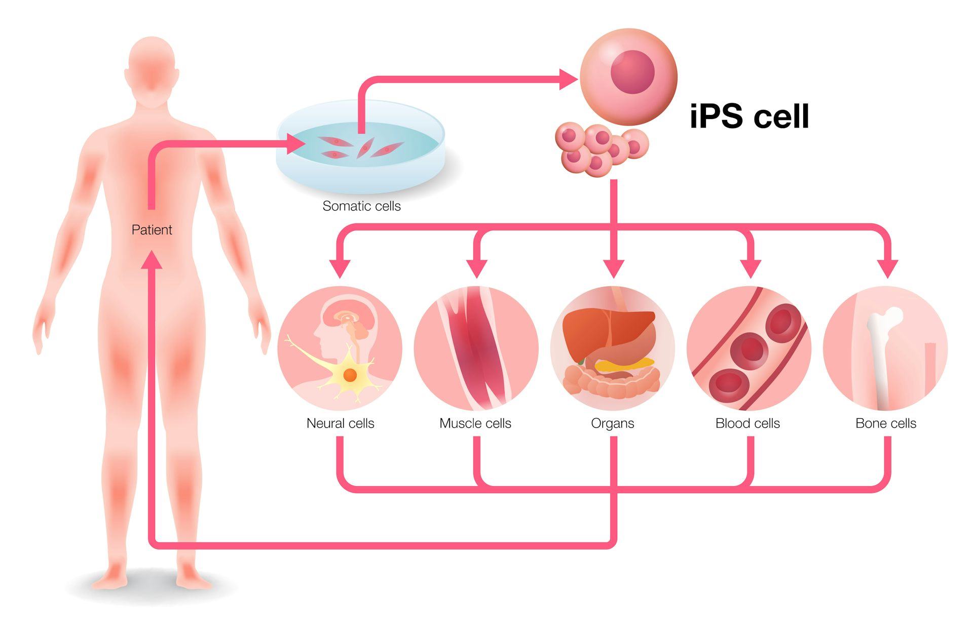
The utility of autologous induced pluripotent stem cell (iPSC) therapies for tissue regeneration depends on reliable production of immunologically silent functional iPSC derivatives. However, rejection of autologous iPSC-derived cells has been reported, although the mechanism underlying rejection is largely unknown. We hypothesized that de novo mutations in mitochondrial DNA (mtDNA), which has far less reliable repair mechanisms than chromosomal DNA, might produce neoantigens capable of eliciting immune recognition and rejection. Here we present evidence in mice and humans that nonsynonymous mtDNA mutations can arise and become enriched during reprogramming to the iPSC stage, long-term culture and differentiation into target cells. These mtDNA mutations encode neoantigens that provoke an immune response that is highly specific and dependent on the host major histocompatibility complex genotype. Our results reveal that autologous iPSCs and their derivatives are not inherently immunologically inert for autologous transplantation and suggest that iPSC-derived products should be screened for mtDNA mutations.


iPSC-derived grafts can be immunogenic and rejected by the host even though they are derived from host cells. Several factors relating to the reprogramming of somatic cells, expansion of iPSCs in culture and differentiation of iPSCs into tissue cells are thought to contribute. Suppression1 and overexpression2 of pluripotency factors are known to establish denovo antigenicity that diminishes with differentiation1,3,4. However, differentiation of iPSCs can lead to the expression of immunogenic antigens not usually expressed in corresponding somatic cells causing rejection.5 In addition, muta-tions acquired during reprogramming and expansion may generate mutant proteins that can act as neoantigens.Mutation rates during reprogramming have been reported to be up to ninefold higher than the background mutation rate in culture.6 Furthermore, the mutation rate for mtDNA is 10- to 20-fold higher than that of nuclear DNA7–9. Both mutated and wild-type mtDNA can coexist in the same cell, a phenomenon called heteroplasmy10. Nonsynonymous mtDNA mutations can impact both the function of proteins11 and its antigenicity. mtDNA-encoded mitochondrial minor antigens12 have been described as transplant barriers13, and our group has shown that individual single nucleotide polymor-phisms (SNPs) are sufficient to create immunogenic neoantigens14. However, the extent to which neoantigenic SNP enrichments affect autologous immune responses are not known. Here we sought to characterize the immunogenicity of mtDNA SNPs and assess their immunologic relevance for iPSC-based regenerative therapies.First, we assessed the sensitivity and specificity of the mouse immune system to respond to isolated mtDNA SNPs. Using the tech-nique of somatic cell nucleus transfer, embryonic stem cells (ESCs) with BALB/c (B/c) nuclear DNA and C57BL/6 (B6) mtDNA were generated (these cells are referred to as NT-ESCs throughout). In comparison to B/c, these NT-ESCs showed only two homoplasmic nonsynonymous SNPs in the mt-Co3 and mt-Cytb genes, and gen-erate cytochrome C oxidase III (Co3) or cytochrome b (Cytb) pro-teins with one amino acid substitution each (Supplementary Table 1). NT-ESCs were used for immunization against these two epitopes in B/c mice (Fig. 1a). To confirm that mitochondrial proteins with a single amino acid substitution can function as neoantigens, B/c fibroblasts were transfected to transiently overexpress either the B/c or B6 forms of Co3 or Cytb (Fig. 1a,b). Splenocytes recovered after 5 d were used for enzyme-linked immunospot (ELISpot) assays against either of the four fibroblast preparations. ELISpot assays for interferon-γ (IFN-γ) and interleukin-4 (IL-4) were performed.


Spot frequencies are presented to directly compare the immune response against the allogeneic B6 protein with that of the syngeneic B/c protein (Fig. 1c). Only the two fibroblasts overexpressing alloge-neic B6 protein evoked a substantial Tcell response. When trans-planted into B/c recipients (Fig. 1d), only those two fibroblast grafts underwent rejection (Fig. 1e,g) during the time period of protein overexpression (Fig. 1f,h). Mass cytometry of splenocytes recovered after 5 d revealed remodeling of the T and B cell compartments as Both high-passage iPSC lines from B6 and FVB revealed potential neoantigens. Single-cell sequencing of B6 cells showed that the heteroplasmies obtained in bulk sequencing accurately reflected the average of more heteroge-neous heteroplasmies in individual cells (Supplementary Table 9).The MHC binding affinities of the B6 neoantigen candidates were modeled using in silico prediction (Supplementary Fig. 8c and Supplementary Table 10), which showed that the mutant SNP in mt-Co1 generated peptides with decent MHC binding affinities whereas the affinity of the mutant Nd3 peptide was very low. Twenty-residue oligomers were synthesized from a sequence carrying the neoanti-genic or reference SNP (Supplementary Table 11). Mice were immu-nized with low-passage or high-passage iPSCs and splenocytes were challenged with 20-residue oligomers 5 d later (Supplementary Fig. 8d–f). As predicted, in B6 mice we observed IFN-γ and IL-4 responses against the Co1 neoantigen but not the mutant Nd3. Immunogenicity was also demonstrated for the 20-residue oligomer of the Atp8 neoantigen in FVB (Supplementary Fig. 8g–i).B6 (Fig. 3c–e) and FVB mice (Supplementary Fig. 9) then received subcutaneous grafts of either low-passage or high-passage iPSCs to assess whether the neoantigens could provoke an immune response and diminish cell survival invivo. Varying cell numbers were used, as the survival and teratoma formation of pluripotent cell grafts depends on the overall cell load16. Across graft sizes, we observed an increased immune response and a correspondingly reduced survival of high-passage iPSC grafts.


... / ...


F O R   T H E R E S T   O F   T H E   R E S E A R C H   A R T I C L E,    V I S I T    T H E   S O U R C E (.ePDF)


#2 Engadin

  • Topic Starter
  • Guest
  • 198 posts
  • 580
  • Location:Madrid
  • NO

Posted 02 September 2019 - 04:21 PM

.


Another approach, not so scientifically deep:


S O U R C E :   TheConversation

T I T L E :    Stem cells could regenerate organs – but only if the body won’t reject them


Many of the most common diseases, like heart failure, liver failure, Type 1 diabetes and Parkinson’s disease, occur when cells or whole organs fail to do their job. Wouldn’t it be fantastic if it were possible to replace cells in these defunct organs? That is exactly what physician-scientists in the field of regenerative medicine are trying to do.


I am a surgeon and stem cell scientist and am interested in regenerating failing organs with stem cells – because for many diseases we don’t have good treatment options yet.


In a recent paper, my colleagues and I figured out why stem cells derived from a patient’s own tissue are sometimes rejected by their own immune systems. We also developed a solution that we think may solve the problem: stem cells that are stripped of their immune features and can’t trigger rejection.


The search for the ideal starter cell


A few years ago a breakthrough occurred that many scientists believed would help fast-track the goal of regenerating organs. That was the identification of proteins that turn on genes that allowed researchers to reprogram adult cells. These proteins transformed cells back into their embryonic-like stem cell state. This gives them the capacity to turn into almost any cell type – like liver or heart or any other cell of interest.


These stem cells can theoretically be used as an inexhaustible source for cells. Scientists believed these cell products could be used to restore the functions of organs and treat diseases. However, regenerating cells and organs from a patient’s own cells and then returning them to that same patient turned out to be trickier than expected.


Researchers are still debating what is the ideal starting cell type for regenerative medicine. The cells required for these therapies can be grown in bioreactors in the lab. But for cell therapies to succeed, the biggest hurdle we have to overcome is immune rejection.


Like transplanted organs, transplanted cells are susceptible to attacks by the recipient’s immune system. Any cells generated from another individual have different proteins on their surface, called tissue antigens, that tag them as “foreign.”


Once tagged, white blood cells, which defend the body against bacteria, viruses and foreign tissue, target these therapeutic cells for destruction. Physicians use high-dose immunosuppressive drugs to silence this immune response so that patients can tolerate a transplanted organ. But these drugs have significant side effects.


To create cells for use in regenerative medicine, scientists envision large-scale collections of stem cells with diverse characteristics and specific tissue antigens. Then just as blood types can be matched, these cataloged stem cells could be matched to the recipient to avoid the patient’s immune system from rejecting these new cells.


One day, hospitals may have enough cell lines to match patients with stem cells based on tissue types. Whether enough cell lines can be banked to serve the wider patient population and whether this strategy will prevent immune responses is yet to be seen.


file-20190819-123720-1riq98j.jpg


Adult cells are removed from patients, transformed into so-called induced pluripotent stem cells and then, using various chemicals, the cells are made to differentiate into different tissue types. Ideally these are then transplanted into the same patient to fix their damaged tissues.


Hurdles for using a patient’s own stem cells


Stem cells generated from a patient’s own cells – called autologous stem cells – are currently believed to be the most promising strategy for circumventing immune rejection. Autologous stem cells are generated directly from the patient seeking treatment and need to be differentiated into the cell type that needs to be replaced. Since the cells carry the same tissue antigens as the patient, they are tagged as “self,” and immunologists believe these cells are accepted by the immune system.


However, this notion may not be correct. In a previous study, our lab had revealed that minor genetic mutations in the DNA carried by a special part of the cell’s DNA, the mitochondrial DNA, can trigger an immune response.


Mitochondria are small structures inside cells that carry their own set of genes that are responsible for generating energy for the cell. Because every cell has many mitochondria, they carry many copies of the mitochondrial DNA. Spontaneous changes in mitochondrial genes, called mutations, alter the shape of the proteins they encode. These mutated proteins, which we call “neoantigens,” re-tag the cells as “foreign,” alert the immune system and target the stem cells for destruction.


Cells that lack immune features may be the solution


Our latest study reveals that neoantigens can spontaneously occur in a patient’s own cells. This renders them susceptible to rejection when used as part of stem cell-based treatment. We showed in mice and humans that minor changes in the mitochondrial DNA can occur when the patient’s cells are being reprogrammed into stem cells so that they can produce different types of cells. This can also happen while the cells are multiplying in plates or bioreactors outside of the body, giving rise to neoantigens.


The likelihood of neoantigens arising increases with the time it takes to manufacture a particular type of cell. If white blood cells recognize neoantigens after injecting the cells back into the animal or human, they may trigger a strong immune response leading to tissue rejection.


Neoantigens can thus jeopardize the whole strategy of autologous cell transplantation. So to use this form of cell transplantation, it may be necessary to test all cell products for mutations in the mitochondrial DNA.


To dodge the immune system and make regenerative stem cell therapies widely available to the general public, our lab aims to engineer stem cells lacking any immune features.


Modern gene editing tools now allow us to make very specific edits and create engineered cell products without any tissue type tags. We recently published our early success with both edited mouse and human stem cells, which survived after transplantation into different mouse models with different tissue types. This was the first report of “universal cells” that completely circumvented rejection by a foreign immune system. We believe this concept could lead to the manufacturing of universal cell products for all patients and has the potential to transform health care.


.


Edited by Engadin, 02 September 2019 - 04:24 PM.


Click HERE to rent this BIOSCIENCE adspot to support LongeCity (this will replace the google ad above).


Also tagged with one or more of these keywords: stem cell research, transplant immunology

0 user(s) are reading this topic

0 members, 0 guests, 0 anonymous users