


Bioconjugated Carbon Dots for Delivery of siTnfα to Enhance Chondrogenesis of Mesenchymal Stem Cells by Suppression of Inflammation
S O U R C E & F U L L T E X T : Stem Cells Traslational Medicine
ABSTRACT
Although a promising strategy, the mesenchymal stem cell (MSC)‐based therapy of cartilage defects is sometimes accompanied with chronic inflammation during the remodeling status, which may hinder cartilage regeneration. During this process, the inflammatory cytokine tumor necrosis factor α (TNFα) plays an important role and may be a potential target. In this study, we investigated the effect of Tnfα RNA interference by introducing a functional and highly safe carbon dot (CD)‐SMCC nanovector synthesized by bioconjugation of CDs with a protein crosslinker, sulfosuccinimidyl‐4‐(N‐maleimidomethyl) cyclohexane‐1‐carboxylate (sulfo‐SMCC), as the vehicle of the silenced TNFα (siTnfα) on chondrogenesis of MSCs. The results showed that CD‐SMCC displayed intense fluorescence with well‐dispersed and positively charged properties, which favored effective binding and delivering of siTnfα into the MSCs. CD‐SMCC‐siTnfαnanoformula also exhibited considerably high transfection efficiency and nearly no cytotoxicity, which is preferred over commercial polyethyleneimine. Interference of Tnfαby CD‐SMCC‐siTnfα markedly promoted the chondrogenesis of MSCs, as indicated by upregulating cartilage‐specific markers. Furthermore, in vivo exploration indicated that CD‐SMCC‐siTnfα transfected MSCs accelerated cartilage regeneration. In conclusion, this study demonstrated that in combination with the novel CD‐SMCC nanovector, targeting Tnfα may facilitate stem cell‐based therapy of cartilage defects. Stem Cells Translational Medicine 2019;8:724&736
Significance Statement
This study developed a functional and highly safe CD‐SMCC nanovector synthesized by bioconjugation of carbon dots (CDs) with a protein crosslinker, sulfosuccinimidyl‐4‐(N‐maleimidomethyl) cyclohexane‐1‐carboxylate (sulfo‐SMCC), which displayed intense fluorescence with well dispersed and positive‐charged properties favorable for effective binding and delivering of small interfering RNA. It was found that interference of Tnfα by CD‐SMCC‐siTnfα markedly promoted the chondrogenesis of MSCs, which is preferred over commercial polyethyleneimine. The study demonstrated that Tnfα may be a potential target and, in combination with the novel CD‐SMCC nanovector, is promising for stem cell‐based therapy of cartilage defects.
Introduction
Mesenchymal stem cells (MSCs) have recently attracted much attention as sources for the clinical therapy of a variety of diseases 1, 2, including cartilage injury, due to their ease of expansion and wide range of functions 3, 4. Recruited MSCs can be specifically induced along the chondrogenic lineage in the presence of specific growth factors, molecules, and epigenetic modifications. However, the immune modulation capability of MSCs is somehow suppressed by chronic inflammation 5 during regeneration 6, which impairs the chondrogenesis of MSCs and restrains the major constituent of the cartilage matrix (type II collagen), thus remaining a challenge for MSC therapy of cartilage defects.
Notably, tumor necrosis factor‐α (TNFα) is one of the proinflammatory cytokines mediating local inflammatory processes in the joint and has an inhibitory effect on chondrogenesis. Elevated TNFα is detected in synovial tissue and fluid during the cartilage destruction cascade 7, which suppresses the synthesis of cartilage matrix (e.g., aggrecan and type II collagen) 8, 9. The potential mechanism may involve TNFα that activates a transcription factor, NF‐κB, which in turn inhibits the synthesis of SOX9, which is a crucial transcription factor required for chondrogenic differentiation 10, 11. TNFα also induces cell apoptosis in chondrocytes 12 and inhibits the migration of chondrogenic progenitor cells from nonfibrillated osteoarthritic cartilage 13. Interference of TNFα obviously eases cartilage degeneration and promotes cartilage regeneration in post‐traumatic arthritis 14, 15. These findings suggest that TNFα may influence chondrogenic differentiation and may be one of the potential targets for MSC therapy. mRNA silencing of TNFα in MSCs by using small interfering RNA (siRNA)‐mediated RNA interference (RNAi) may enhance chondrogenesis.
Successful gene delivery requires that the vehicles have favorable biocompatibility to avoid the elicitation of an adverse host immune response, as well as high transfection efficiency. Among the various vehicles, nanoparticles have attracted the most attention due to their unique properties, including as biosafety, flexibility, ease of formulation, and high delivery efficiency 16. Among the various carriers, quantum dots (QDs) and carbon dot (CD)‐based gene delivery systems are two of the most widely used fluorescent delivery systems 17, 18. In a previous study, we reported on QD nanovectors modified by sulfosuccinimidyl‐4‐(N‐maleimidomethyl) cyclohexane‐1‐carboxylate (sulfo‐SMCC) for gene delivery and gene targeting in MSCs 19. Although efficient in gene silencing, the heavy metal toxicity of QDs hinders its application to gene therapy in vivo 20. Another fluorescent nanomaterial with both favorable biocompatibility and monodispersity, CDs have been proven to be excellent and efficient gene delivery vectors 21 and may replace QDs as the leading carrier in gene transfer. However, the gene may not bind closely to CDs without optimized surface modification 22. A previous study from our laboratory demonstrated that QDs bioconjugated with sulfo‐SMCC can form a stable thioether bond 19, which retained the integrity of siRNA 23 and greatly increased the transfection efficiency 24. Sulfo‐SMCC contains N‐hydroxysuccinimide ester and maleimide groups, allowing stable covalent conjugation of amine‐containing and sulfhydryl‐containing molecules 25. This may shed light on the modification of CDs.
Based on our previous study, we synthesized CD‐SMCC by bioconjugation of CDs with SMCC for the gene transfer of silenced TNFα (siTnfα) in MSCs to assess the effects of siTnfα on chondrogenic differentiation, with the intention of enhancing chondrogenesis of MSCs and further cartilage regeneration in vivo (Fig. 1). Polyethyleneimine (PEI) was used as the control. This study may provide new insight into cartilage repair.
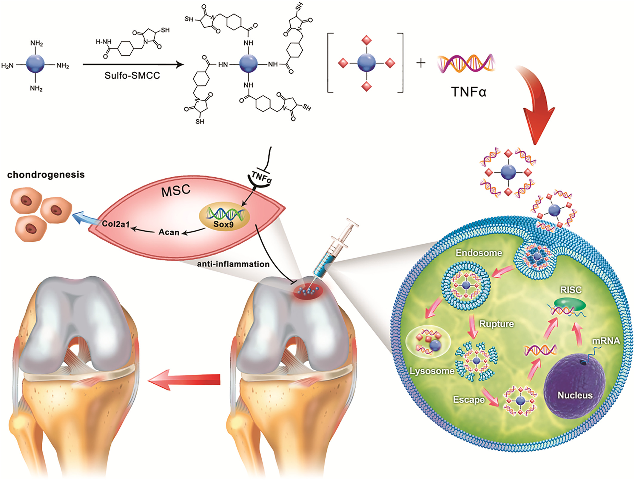
FIGURE 1. Schematic illustration of the formation of carbon dot (CD)‐succinimidyl‐4‐(N‐maleimidomethyl) cyclohexane‐1‐carboxylate‐silenced TNFα complexes and the CD‐based nanocarrier for gene delivery and real‐time monitoring of cellular trafficking in vitro and in vivo for enhancing cartilage repair.
..../....
.
Edited by Engadin, 15 July 2019 - 12:45 PM.


