


A new publication in iScience has described a novel way in which heart tissue can be encouraged to accept a gene therapy by using ultrasound to create cavitation bubbles.
A little-known target and a new delivery vector
This paper begins with a discussion of S-adenosylhomocysteine (SAH), a compound that, in excess, is associated with an increase in cardiovascular risk [1]. This compound is converted to other molecules by SAH hydrolase (SAHH), and inhibiting SAHH has been found to increase oxidative stress, inflammation, and diabetes risk in a mouse model [2].
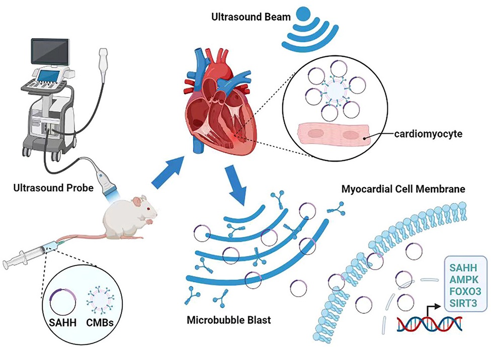
Therefore, these researchers decided that upregulating SAHH may be a viable approach for reducing cardiovascular risk. However, getting the right cells to produce SAHH was previously impossible, which is why this team has chosen a novel approach: ultrasound-targeted microbubble destruction (UTMD). This technology uses low-intensity ultrasound to allow microbubbles containing the desired genes to enter heart tissue, where they rupture and release their payloads [3].
Beneficial effects in rats
Through the combination of a specifically poor diet and a known toxin, these researchers induced diabetic cardiomyopathy (DCM) in rats. Compared to a control group, these rats expressed significantly less SAHH, as confirmed by standard Western blots and by gene expression analysis. As expected, oxidative stress in the DCM rats was also increased.
After confirming that their SAHH-based treatment worked in a cellular culture, making sure that id was not dangerous to cells and that it did indeed increase SAHH in the targeted cells, the researchers then administered it to the DCM rats. They first made sure that the treatment was actually targeted towards only cardiac tissue; analyzing other tissues, such as the spleen, the lung, and the kidney, they found no evidence of increased SAHH.
Instead, they found that their treatment was reaching the targeted cells. The effect of the treatment was moderate but statistically significant. SAHH expression in the treatment group was restored about halfway to the level of the non-DCM control group. Cellular death by apoptosis was slightly reduced in the treatment group compared to the DCM control group, but not close to the level of the non-DCM control group; this was confirmed by a gene expression analysis, which found that the related apoptotic signals were significantly diminished in the treatment group.
Benefits to oxidative stress and to heart tissue
Oxidative stress was also found to be significantly decreased in the treatment group. The AMPK pathway responsible for protecting cells against this stress was upregulated, and reactive oxygen species were decreased by the treatment.
An examination of the rats’ hearts one month after the treatment found that there were far fewer broken fibers in the treatment group. Collagen deposition, another marker of damage, was moderately diminished.
If these positive results can be replicated in human beings, an entirely new method of treating diabetes-related heart failure may be at hand. However, further experiments along with clinical trials will need to be conducted to determine if this approach can be translated to medical practice.
Literature
[1] Kerins, D. M., Koury, M. J., Capdevila, A., Rana, S., & Wagner, C. (2001). Plasma S-adenosylhomocysteine is a more sensitive indicator of cardiovascular disease than plasma homocysteine. The American journal of clinical nutrition, 74(6), 723-729.
[2] Dai, X., Liao, R., Liu, C., Liu, S., Huang, H., Liu, J., … & Xiao, Y. (2021). Epigenetic regulation of TXNIP-mediated oxidative stress and NLRP3 inflammasome activation contributes to SAHH inhibition-aggravated diabetic nephropathy. Redox Biology, 45, 102033.
[3] Qin, X., Cai, P., Liu, C., Chen, K., Jiang, X., Chen, W., … & Tian, H. (2023). Cardioprotective effect of ultrasound‐targeted destruction of Sirt3‐loaded cationic microbubbles in a large animal model of pathological cardiac hypertrophy. Acta Biomaterialia, 164, 604-625.
View the article at lifespan.io


