


Researchers have found that altering a growth hormone receptor in the brain adipose tissue of aged male mice slows their mental aging and allows them to perform far better on cognitive tests.
Growth signaling is not necessarily good
The axis of growth hormone and insulin-like growth factor 1 (IGF-1) is well-known in aging, and the relationship between this regulator and brain aging has been previously documented [1]. Interestingly, while circulating growth hormone and IGF-1 levels decline with aging [2], suppressing this signaling extends lifespan [3], and mice with reduced levels of this signaling perform better on cognitive tests [4]; this also occurs when the mice express an agonist that suppresses it [5].
The researchers of this study focused on its effects on fatty (adipose) tissue, which is metabolically active and secretes factors that affect other systems [6], including the brain [7]. While previous work has discovered that adipose-specific growth hormone knockout (Ad-GHRKO) mice have better insulin sensitivity and longer lives [8], how well these mice perform on cognitive tests had not been previously measured.
Benefits for neural function and inflammation
The researchers first directly examined the brains of these mice, comparing 18- to 24-month-old Ad-GHRKO mice to controls. The modified mice were more neurally active overall and had less neuronal loss in the dentate gyrus, the part of the hippocampus responsible for forming new memories. This was accompanied by an increase in synapse formation and a decrease in neuroinflammation: there were decreases in the inflammatory factors IL-6 and TNF-α along with an increase in the anti-inflammatory factor IL-10.
There was also a reduction in cellular senescence. The modified mice had significantly less of the senescence marker SA-β-gal throughout their brains, including the amyglada, the dentate gyrus, and the cortex. They also had significantly less tau phosphorylation, an age-related protein alteration that contributes to cognitive decline and, in humans, is a sign of Alzheimer’s disease.
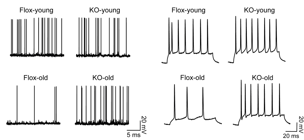
The excitability of neurons declines with age, and here, too, knocking out growth hormone in the adipose tissue proved beneficial; the aged modified mice fired their neurons much more like younger mice did, while aged controls had stark reductions in neural firing frequency.
Stark benefits on cognitive tests
The researchers then turned to four standard cognitive tests: the novel object recognition test, which shows that the mice can discriminate between familiar and unfamiliar things; the Y-maze tests, which tests for exploration ability; the Morris water maze test, which tests memory and navigation behavior; and a floor shock test, which tests memory in adversive conditioning. On all four of these tests, the aged modified mice performed almost exactly like their younger counterparts, while the aged controls performed far worse; this was in spite of the modification not noticeably affecting the older mice’s physical ability.
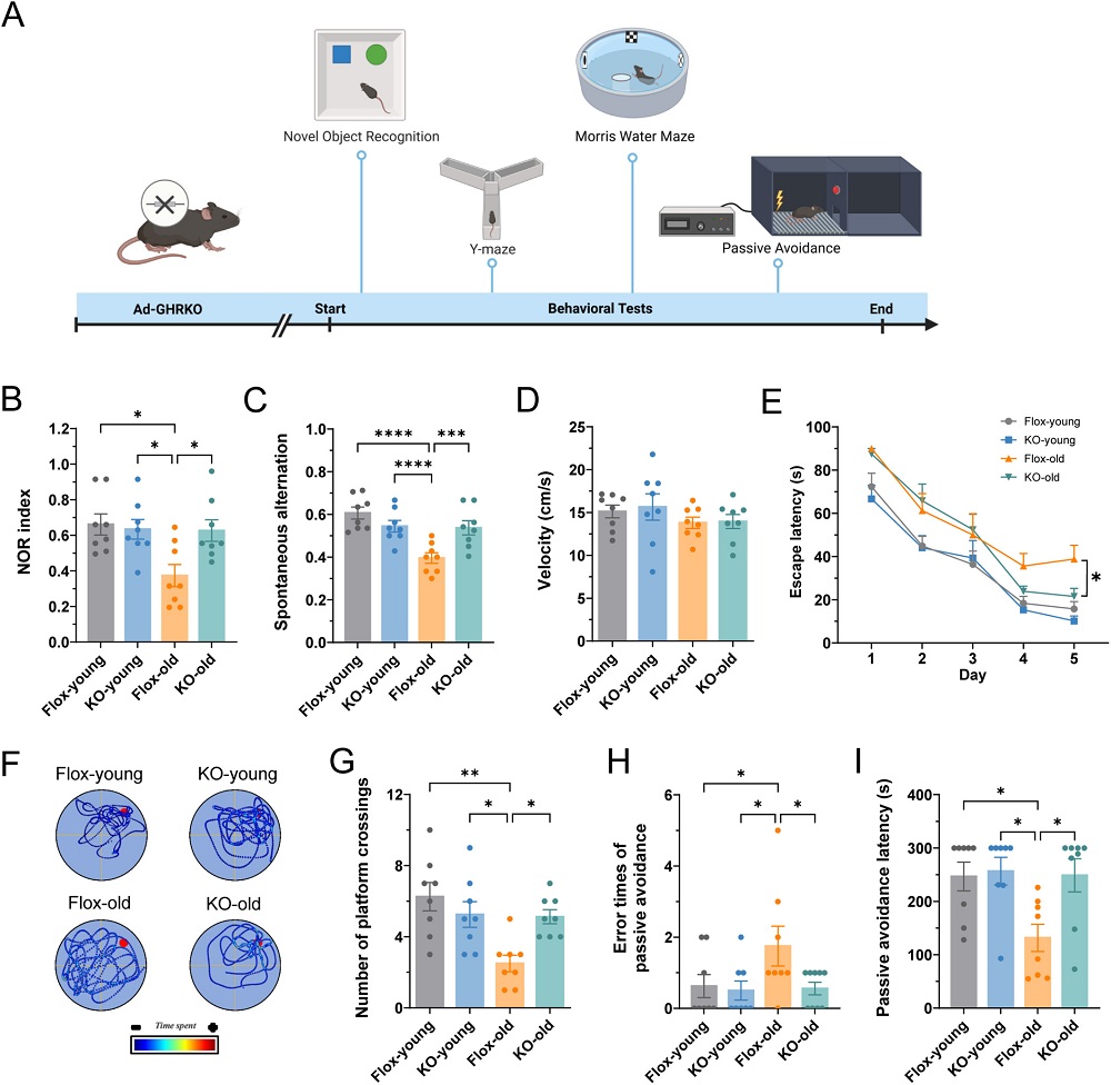
According to the authors, “this study provides evidence that adipose tissue acts as a key peripheral regulator of brain aging.” While the number and power of cognitive and biochemical benefits that arose from this modification are striking, these experiments were performed on a single-sex group of genetically altered mice. Aapplying these findings to wild-type animals, and then human beings, is a challenge of its own.
Literature
[1] Ashpole, N. M., Sanders, J. E., Hodges, E. L., Yan, H., & Sonntag, W. E. (2015). Growth hormone, insulin-like growth factor-1 and the aging brain. Experimental gerontology, 68, 76-81.
[2] Liu, H., Bravata, D. M., Olkin, I., Nayak, S., Roberts, B., Garber, A. M., & Hoffman, A. R. (2007). Systematic review: the safety and efficacy of growth hormone in the healthy elderly. Annals of internal medicine, 146(2), 104-115.
[3] Bartke, A. (2008). Growth hormone and aging: a challenging controversy. Clinical interventions in aging, 3(4), 659-665.
[4] Kinney-Forshee, B. A., Kinney, N. E., Steger, R. W., & Bartke, A. (2004). Could a deficiency in growth hormone signaling be beneficial to the aging brain?. Physiology & behavior, 80(5), 589-594.
[5] Basu, A., McFarlane, H. G., & Kopchick, J. J. (2017). Spatial learning and memory in male mice with altered growth hormone action. Hormones and Behavior, 93, 18-30.
[6] Booth, A., Magnuson, A., Fouts, J., & Foster, M. T. (2016). Adipose tissue: an endocrine organ playing a role in metabolic regulation. Hormone molecular biology and clinical investigation, 26(1), 25-42.
[7] Letra, L., & Santana, I. (2017). The influence of adipose tissue on brain development, cognition, and risk of neurodegenerative disorders. Obesity and Brain Function, 151-161.
[8] List, E. O., Berryman, D. E., Slyby, J., Duran-Ortiz, S., Funk, K., Bisset, E. S., … & Kopchick, J. J. (2022). Disruption of growth hormone receptor in adipocytes improves insulin sensitivity and lifespan in mice. Endocrinology, 163(10), bqac129.
View the article at lifespan.io


