






Researchers have discovered that BNIP3, which regulates mitochondrial maintenance, is increased with aging, harming the heart’s ability to process energy. Mitochondrial transplantation was found to be effective against this increase in a mouse model.
The heart needs energy
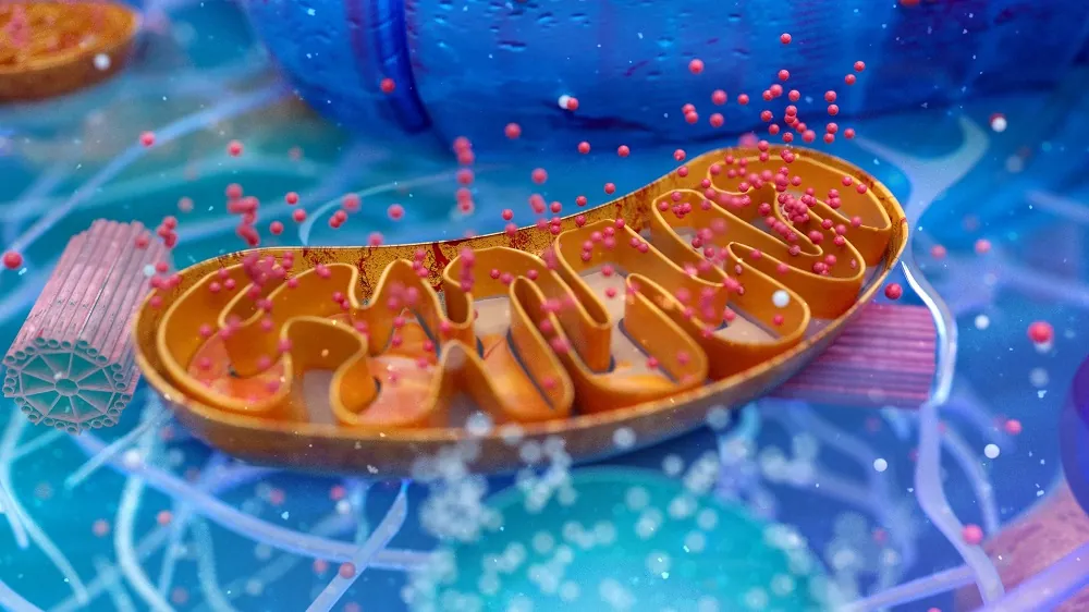
Mitophagy, the process of eliminating damaged mitochondria, is necessary for the proper function of organs and staving off mitochondrial dysfunction [1]. If cells can’t begin or finish the process of mitophagy, the damaged mitochondria accumulate within cells, harming energy production and increasing oxidative stress, particularly in hardworking heart muscle cells (cardiomyocytes) [2], which are the focus of this study.
BNIP3, which is located in the membranes of mitochondria, is activated under stress conditions, spurring the formation of mitophagosomes, which conduct mitophagy, by recruiting LC3B in autophagosomes [3]. Normally, this would be beneficial, but BNIP3 is also linked to cellular death by apoptosis, and excessive BNIP3 is linked to heart failure [4].
We have recently reported on research that uses transplanted cells to donate mitochondria in the brain. This team has previously found that the same can occur in the heart [5], but that previous work did not closely investigate energy metabolism nor did it look at BNIP3.
Overfilling the cellular incinerator
Microscopic examination determined that senescent heart cells have more, not fewer, mitophagosomes than younger cells. LC3B is upregulated as well. When chloroquine, which damages mitochondria, is introduced to younger cells, it stimulates increases in LC3B; however, it does nothing to increase mitophagy in senescent cells. Therefore, the mitophagy process is “functionally saturated”: mitophagy initiation is boosted to its maximum, but the damaged mitochondria are not being destroyed quickly enough to keep up, leading to their accumulation. This is referred to as a blockade of mitophagic flux.
Transplantation of mitochondria from mesenchymal stem cells (MSCs) alleviated some of this dysfunction in mice that had been artificially aged through the administration of doxorubicin, which causes cells to become senescent. Compared to a control group that had only been aged in this way, aged mice given these mitochondria experienced less senescence as measured by SA-β-gal, better heart function according to measurements of volume and flow, and reductions of age-related mitophagosome accumulation.
Unsurprisingly, this aging was found to be connected to an increase in murine Bnip3 along with other senescence-related genes, and this result was confirmed by data derived from naturally aged mice as well as data from human cells. The PINK1/Parkin mitophagy pathway was not affected. In the researchers’ mouse model, murine Bnip3 expression was reduced by mitochondrial transplants.
BNIP3 needs to be regulated
Further work with human cardiomyocytes confirmed that BNIP3 is a cause rather than a downstream consequence. Upregulating BNIP3 in these cells led directly to an increase in the senescence marker p16. Creating mice that were both artificially aged and overexpress BNIP3 led to mitochondrial transplantation having no benefit. An examination of LC3B determined that “aberrant BNIP3 expression directly perturbs the process of mitophagy,” meaning that preventing the age-related increase in its overexpression is key to preventing the blockade of mitophagic flux and thus delaying the senescence of heart cells.
Using both animal and human data, the researchers also found that a hypoxia-inducible factor, HIF-3α, is a regulator of BNIP3, which concurs with previous work [6]. Unsurprisingly, HIF-3α was upregulated in aged human and mouse cardiomyocytes. Working with human cardiomyocytes, the researchers found that overexpressing HIF-3α overexpresses BNIP3 as well, and BNIP is depleted when HIF-3α is depleted. Knocking down HIF-3α while directly overexpressing BNIP3 reduces some of BNIP3’s negative effects.
By using CCCP, an inducer of mitochondrial damage, the researchers found that HIF-3α is overexpressed when ATP is scarce. Therefore, at least some of the benefits of mitochondrial transplantation stem from its resulting increases in ATP, which diminish HIF-3α and thus BNIP3, clearing the blockage of mitophagic flux.
However, the biochemical relationship between HIF-3α and ATP scarcity was not determined, and the researchers note that they did not analyze how native mitochondria and transplanted mitochondria interact. Furthermore, this was only a murine and cellular study, and it is not yet clear if this approach is safe for human beings.
Literature
[1] Lin, J., Chen, X., Du, Y., Li, J., Guo, T., & Luo, S. (2024). Mitophagy in cell death regulation: insights into mechanisms and disease implications. Biomolecules, 14(10), 1270.
[2] Xu, X., Pang, Y., & Fan, X. (2025). Mitochondria in oxidative stress, inflammation and aging: from mechanisms to therapeutic advances. Signal transduction and targeted therapy, 10(1), 190.
[3] Lu, Y., Li, Z., Zhang, S., Zhang, T., Liu, Y., & Zhang, L. (2023). Cellular mitophagy: mechanism, roles in diseases and small molecule pharmacological regulation. Theranostics, 13(2), 736.
[4] Wu, Q. Q., Yao, Q., Hu, T. T., Wan, Y., Xie, Q. W., Zhao, J. H., … & Tang, Q. Z. (2022). Tax1 banding protein 1 exacerbates heart failure in mice by activating ITCH-P73-BNIP3-mediated cardiomyocyte apoptosis. Acta Pharmacologica Sinica, 43(10), 2562-2572.
[5] Jin, N., Zhang, M., Zhou, L., Jin, S., Cheng, H., Li, X., … & Xie, J. (2024). Mitochondria transplantation alleviates cardiomyocytes apoptosis through inhibiting AMPKα‐mTOR mediated excessive autophagy. The FASEB Journal, 38(10), e23655.
[6] Huang, L., Wang, L., Yuan, D., Xu, Y., Wang, Y., Yao, K., … & Liu, D. (2025). Overexpression of BNIP3 in renal carcinoma cells can promote apoptosis of renal carcinoma cells through HIF-1α-BNIP3-mediated autophagy. Frontiers in Oncology, 15, 1614378.
View the article at lifespan.io


