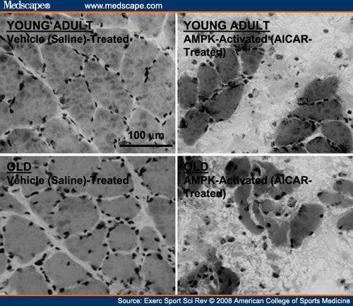
Gradual basal skeletal muscle atrophy with age (sarcopenia) is perhaps even more clinically significant than diminished overload-induced growth. The fact that AMPK activity displays a five-fold increase with age in resting fast-twitch, but not slow-twitch, muscle (Figure 1)[25] led us to speculate that AMPK may partly underlie basal fast-twitch specific atrophy, an effect not testable by acute AMPK activation studies. Thus, we continuously activated AMPK in the resting muscles of young and old rats for 7 d (AICAR perfusion via osmotic pump and catheter). Importantly, we used slow-twitch soleus muscles for this experiment, hypothesizing that aged slow-twitch muscle (previously displaying neither elevated AMPK activity nor atrophy with age (Figure 1)[25] would exhibit sarcopenia-like properties if AMPK were chronically activated similar to aged fast-twitch muscle. As hypothesized, continuous AMPK activation elicited significant fiber atrophy in both young adult and old muscles (Figure 5). Moreover, an additional unexpected finding of continuously activating AMPK in resting muscle was a high frequency of fiber death (Figure 5).
 (Enlarge Image) Figure 5. One week of continuous 5′-AMP-activated protein kinase (AMPK) activation causes fiber atrophy and death in slow-twitch soleus muscles of young adult (YA; 8 mo) and old (O; 30 mo) Fisher344 × Brown Norway F1 hybrid rats. Muscles were locally and continuously perfused (osmotic pump and catheter) with either 5-aminoimidazole-4-carboxamide-1-beta-D-ribofuranoside (AICAR; an AMPK activator) at 0.5 mg·h-1 or vehicle (saline) during the 1-wk period. Fiber cross-sectional area was significantly (P ≤0.05) reduced in both age groups (by 25% in YA muscles and by 24% in O muscles; n = 3 per group). Images are representative muscle cross-sections stained with hematoxylin and eosin.
[ CLOSE WINDOW ]

Figure 5.
One week of continuous 5′-AMP-activated protein kinase (AMPK) activation causes fiber atrophy and death in slow-twitch soleus muscles of young adult (YA; 8 mo) and old (O; 30 mo) Fisher344 × Brown Norway F1 hybrid rats. Muscles were locally and continuously perfused (osmotic pump and catheter) with either 5-aminoimidazole-4-carboxamide-1-beta-D-ribofuranoside (AICAR; an AMPK activator) at 0.5 mg·h-1 or vehicle (saline) during the 1-wk period. Fiber cross-sectional area was significantly (P ≤0.05) reduced in both age groups (by 25% in YA muscles and by 24% in O muscles; n = 3 per group). Images are representative muscle cross-sections stained with hematoxylin and eosin. The fiber atrophy induced by AMPK activation (Figure 5) agrees with findings that cultured myotube diameter is reduced under conditions in which AMPK activity is up-regulated[1] and supports the possibility that chronically elevated AMPK activity may contribute to basal fast-twitch fiber atrophy with age (sarcopenia). Suppression of muscle protein translation and synthesis[4,24] may be one mechanism by which AMPK exerts this effect; however, it is highly doubtful that solely suppressing protein synthesis would elicit the striking fiber atrophy and death observed in just 7 d (Figure 5). Thus, activation of protein degradation pathways is also a likely possibility. As previously mentioned, AMPK is known to stimulate muscle-specific lysosomal and proteasomal gene expression (FOXO, Atrogin-1, and MuRF1) as well as myofibrillar protein degradation.[15,17] The increased FOXO3A and MuRF1 mRNA expression and increased myofibrillar degradation with age in resting human muscle of mixed fiber types[21,28] may therefore result in part from elevated AMPK activity in the fast-twitch population. Interestingly, some have observed elevated rates of mixed[10] and myofibrillar[13] protein synthesis, along with higher S6K activation,[13] in aged rat fast-twitch muscle. Such a scenario would indicate a futile increase in protein synthesis rate in the face of an even faster degradation rate in these muscles, although still others have conversely observed lower basal protein synthesis in aged muscle.[29] Regardless of protein synthesis differences, an elevated protein degradation rate in aged fast-twitch muscle fibers likely plays a role in subtly, and over a long period of time, altering the synthesis-degradation balance toward gradual atrophy. We propose that chronically elevated AMPK activity may play a role in this phenomenon.
The possibility that continuously elevated AMPK activity affects the balance of myonuclear addition versus nuclear apoptosis in aged fast-twitch muscle is intriguing. Unfortunately, few data exist concerning AMPK's potential involvement in either phenomenon in skeletal muscle. Certainly, suppression of basal myonuclear addition cannot account for the rapidity with which AICAR induced fiber atrophy and death (Figure 5), and whether these effects were due to apoptotic mechanisms and/or more general fiber necrosis remains to be determined. However, AMPK does seem to play a key role in apoptotic signaling in mononucleated muscle cell types, including smooth muscle cells, cardiac myocytes, and immortalized (C2C12) skeletal myoblasts.[8,18] It is currently unclear whether AMPK is proapoptotic or antiapoptotic in these cells because there is somewhat equivocal evidence to support both.
In contrast to mononucleated cells, apoptosis in mature multinucleated skeletal muscle fibers does not necessarily mean fiber death but is more associated with fiber atrophy as the myonuclear domain volume remains relatively constant. It is unknown whether nuclear apoptosis leads to protein degradation and fiber atrophy or vice versa (i.e., fiber atrophy leads to nuclear apoptosis, which maintains a constant myonuclear domain volume). As might be expected, the frequency of apoptotic nuclei is well known to be increased in aged atrophying muscle fibers.[5] If AMPK is proapoptotic in mature muscle fibers similar to other muscle cell types under some conditions,[8] then chronically elevated AMPK activity (Figure 1)[25] may play a role in the age-related increase in apoptotic nuclei. In fact, we propose that any potential role of AMPK in aging multinucleated muscle fibers is more likely proapoptotic than antiapoptotic because of the following logic: 1) if myonuclear domain volume is to be kept constant, it is paradoxical to protect against nuclear death while selectively reducing protein synthesis and/or stimulating protein degradation; and 2) the potential antiapoptotic effects of AMPK in mononucleated cells is thought to enhance cell survival during times of brief energy deficit,[8] whereas apoptosis in multinucleated muscle fibers is more associated with atrophy than a risk of cell death (and lost nuclei can be replenished from the myogenic precursor cell pool when energy balance is restored). Moreover, although the extent of atrophy and/or nuclear apoptosis necessary to eventually achieve a "threshold" precipitating complete muscle fiber death is unknown, our data indicate a role for continuous AMPK activation in fiber atrophy and eventual fiber death, not fiber survival (Figure 5). Because fiber atrophy and death are also characteristics of sarcopenic muscle,[29] the potential role of AMPK in mediating apoptosis in aged skeletal muscle fibers, either directly or indirectly (via fiber atrophy and the loss of myonuclear domains) demands further exploration.


