• Log in with Facebook Log in with Twitter Log In with Google      Sign In   
  • Create Account
L onge C ity       Advocacy & Research for Unlimited Lifespans

Photo

Stem Cell Research News


  • Please log in to reply
337 replies to this topic

#241 chubtoad

  • Life Member
  • 976 posts
  • 5
  • Location:Illinois

Posted 11 August 2004 - 10:07 PM

http://www.nature.co.../040809-11.html

UK gives go-ahead to therapeutic cloning

Helen Pilcher
British researchers receive stem-cell licence. 
Therapeutic cloning yields stem cells, which may be used to treat disease.
British scientists have been given the green light to clone human embryos for therapeutic purposes. The move should help researchers to understand how diseases such as Alzheimer's, Parkinson's and diabetes arise, and should speed the development of new therapies.

The Human Fertilisation and Embryology Authority has announced that it is to grant the licence to researchers at the Centre for Life in Newcastle on 12 August. It is the first such licence to be issued in Britain, a country that has one of the strongest regulatory systems in the world on cloning.

"We're absolutely thrilled," says reproductive biologist Alison Murdoch from the Newcastle Fertility Centre at Life. "The potential that this area of research offers is immensely exciting and we are keen to take the work we've done so far to the next level."


#242 DJS

  • Guest
  • 5,798 posts
  • 11
  • Location:Taipei
  • NO

Posted 13 August 2004 - 08:06 AM

Hypothetical scenario: In five years the production of large quantities of ESCs and the refinement of SCNT have been developed. This allows for the transplantion of genetically identitical tissue without the risks associated with immunosupressant therapies.

How would this help with Type I diabetes? Wouldn't the patient still need to be placed on an immunosupressant therapy because the under lying cause of the disease lies in the faulty genetically coded instructions which causes the individual's body to attack "healthy" pancreatic islets? Is there something I'm missing here?

Unless, of course, the technology was proficient enough to constantly rejuvinate a patient's continually depleting beta cells...

Click HERE to rent this BIOSCIENCE adspot to support LongeCity (this will replace the google ad above).

#243 Cyto

  • Topic Starter
  • Guest
  • 1,096 posts
  • 1

Posted 16 August 2004 - 10:05 PM

Stem cell research targets cerebral palsy

Posted Image

"We know that we can get stem cells into the brain and they will turn into brain cells but we really don't know how well they work," says Dr. James E. Carroll, chief of the MCG Section of Pediatric Neurology. "The cells probably do form synapses," he says of connections brain cells make so they can communicate. "But the question is: Will all this integrate into improved function?"

Dr. Carroll is principal investigator on a new grant from the American Heart Association and an existing grant from the National Institutes of Health that are using an animal model of cerebral palsy to identify the most effective way to transplant stem cells and possibly answer that question.

With this latest grant, Dr. Carroll, who also treats patients with cerebral palsy, wants to determine whether transplanted stem cells work best when the cells are injected directly into the brain along with these natural chemicals, called chemokines.

He'll try several direct approaches including injecting chemokines into the injury site first and stem cells second and taking the technically-easier route of injecting both at the same time. To enable this approach, Dr. Carroll will use a virus' ability to infect a cell to get chemokines inside stem cells before they are injected.

He'll also look at the bottom line: whether the motor skills of the animal model are improved following the transplant.

Cerebral palsy, which affects about 500,000 people in the United States, is defined as brain damage that occurs before or during birth. The number of people with the disorder has increased over the last 30 years as more premature babies survive. Its effects run the gamut, from barely detectable to devastating loss of motor control. The causes are diverse as well, including everything from oxygen deprivation during birth to prenatal infections.


#244 Cyto

  • Topic Starter
  • Guest
  • 1,096 posts
  • 1

Posted 18 August 2004 - 11:19 PM

Embryonic Stem Cell Therapy Shows Steady Benefits In Rebuilding Infarcted Heart

Despite improvements in earlier diagnosis and treatment, cardiovascular disease is far and away the leading cause of death in the U.S. and the world. According to the latest posted statistics, heart disease causes 700,000 deaths in the U.S. each year, with the number of adults diagnosed with heart disease exceeding 23 million, or 11.5% of the adult population.

Previously thought of as concentrated in rich countries, ischemic heart and related cerebrovascular diseases alone caused an estimated 17 million or 23.2% of deaths globally in 2000, WHO reported. 

A contributing cause of what some have called a pandemic is that in contrast to many other organs in the body, the heart has only a minimal capability for self-renewal, leaving most current therapies to address symptoms with little hope of rehabilitating the injured heart itself after a heart attack (myocardial infarction).

The regenerative potential of stem cells in relation to the muscle layer of the heart wall (or myocardium) has been recently recognized, but how this might translate into therapeutic uses to repair the heart has been limited.

Findings and discussion

Researchers took a murine embryonic stem cell line and engineered a cell clone to express fluorescent proteins so they could easily identify the location of the “newly” generated cardiac cells. They tested the cardiogenic capacity of the line and collected cells with high potential for becoming cardiomyocytes. Randomly assigned rats that had been induced with myocardial infarction were injected directly into the damaged heart area either with the embryonic stem cells or were subject to a control or “sham” protocol.

Three weeks after therapy, the cardiac contractile function of both groups was tested by echocardiography. Not only was the stem cell-treated group’s left ventricular pumping significantly stronger than the sham-treated group, but the heart beat of the stem-cell group reacted favorably to “stress test,” whereas the sham group showed no significant response at all.

Like all the other parameters, these improvements were maintained over the three-month length of the study.

On pathology, further investigation showed the extent of the positive influence of the stem-cell therapy. First, cardiac cells stayed in the heart, and didn’t spread to the brain, kidney or liver. Microscopy showed that the stem cell-derived cardiomyocytes took on the distinctive striations indicating proper development of contractile apparatus. Stem cell-treated hearts also showed normal cardiac ultrastructure, in contrast to acellular infarct areas of sham-treated hearts.

Finally, the stem cell-treated hearts showed that the wall or muscle had been “rebuilt” compared with the sham-treated hearts which remained “eaten-up”, with a decayed thin look, including formation of aneurysms, associated with a post-heart-attack liability to rupture. Conversely, the stem cell-treated hearts showed no evidence of graft rejection, electrical and/or structural abnormality, sudden cardiac death or tumor formation.

Conclusion and next steps

The researchers conclude that “embryonic stem cells, through differentiation within the host myocardium, can contribute to a stable beneficial outcome on contractile function and ventricular remodeling in the infarcted heart.”

Going a step further, they add that “the stable benefit of embryonic stem cell therapy on myocardial structure and function in this experimental model supports the potential for stem cell-based reparative treatment of myocardial infarction. By regenerating diseased myocardium and promoting cardiac repair, embryonic stem cells provide a unique therapeutic modality that has the potential to reduce the morbidity and mortality of this prevalent heart disease.”

Looking ahead, they noted that issues to be resolved include mechanisms of action, finding the optimum window for therapy, and determining what the long-term effect of such therapy will be.

Editors’ note: A copy of this research paper is available to the media. Members of the media are encouraged to obtain an electronic version and to interview members of the research team. To do so, please contact Mayer Resnick at APS 301.634.7209, cell 301.332.4402 or mresnick@the-aps.org.

The American Physiological Society was founded in 1887 to foster basic and applied bioscience. The Bethesda, Maryland-based society has more than 10,000 members and publishes 14 peer-reviewed journals containing almost 4,000 articles annually.


#245 DJS

  • Guest
  • 5,798 posts
  • 11
  • Location:Taipei
  • NO

Posted 20 August 2004 - 04:27 PM

Is it inappropriate to have Q&A on this thread or does no one have the answers to my questions? Or are my questions misguided, or does no one have the the time at the moment, or.... :)

Somtimes this site doesn't seem very user friendly for new comers and students of biotech. Maybe there could be a Q&A and/or a tutorial thread.

#246 Cyto

  • Topic Starter
  • Guest
  • 1,096 posts
  • 1

Posted 20 August 2004 - 11:15 PM

Heh, sorry about that. Emmm, give me a moment with the questions.

A Q&A...I would be more that happy to be apart of a list of where you can ask questions via PM and then, with consent from both parties, will be posted once an answer is given. Might we pin a all caps topic titled "ASK A BIOTECH QUESTION" or something of the sort.

I will openly admit that if I were PMed the questions I would answer them 100% where-as with a post of questions I am more prone to overlook or forget about.

Click HERE to rent this BIOSCIENCE adspot to support LongeCity (this will replace the google ad above).

#247 Cyto

  • Topic Starter
  • Guest
  • 1,096 posts
  • 1

Posted 21 August 2004 - 12:48 AM

If we were to do a BioPump section of the skin to secrete insulin (epithelial cells near adequate vasculature) it should bypass a need for replacement cells. Wouldn't last as long as stem cell replacement but I do find most articles not willing to talk about the immunosuppression efforts to ensure newly anchored replacements survivability. Nor do I know if we could have epithelial cells adjust insulin production via glucose concentrations...I would need to look at the current understanding of such transduction pathways. I think BioPump research would be a more patient-friendly avenue to pursue.

There are other ideas about this too...

Nanoporous microsystems for islet cell replacement
doi:10.1016/j.addr.2003.11.006

The inadequacy of conventional insulin therapy for the treatment of Type I diabetes has stimulated research on several therapeutic alternatives, including insulin pumps and controlled release systems for insulin. One of the most physiological alternatives to insulin injections is the transplantation of insulin-secreting cells. It is the beta cells of the islets that secrete insulin in response to increasing blood glucose concentrations. Ideally, transplantation of such cells (allografts or xenografts) could restore normoglycemia. However, as with most tissue or cellular transplants, the cellular grafts, particularly xenografts, are subjected to immunorejection in the absence of chronic immunosuppression. Thus, it is of great interest to develop new technologies that may be used for islet cell replacement. This research proposal describes a new approach to cellular delivery based on micro- and nanotechnology. Utilizing this approach, nanoporous biocapsules are bulk and surface micromachined to present uniform and well-controlled pore sizes as small as 7 nm, tailored surface chemistries, and precise microarchitectures, in order to provide immunoisolating microenvironments for cells. Such a design may overcome some of the limitations associated with conventional encapsulation and delivery technologies, including chemical instabilities, material degradation or fracture, and broad membrane pore sizes.


Ill take a look at the transduction pathways to see how complex a system we are talking about and post again.

#248 bradbury

  • Guest
  • 2 posts
  • 0

Posted 21 August 2004 - 09:24 AM

Speaking to some of Don's comments... Anytime (in the current medical paradigm) you are talking about growing large numbers of personal stem cells you are talking about a relatively expensive therapy -- at least until we have built up a large infrastructure capable of doing this on a mass-production basis. So if one does not deal with the underlying problem of the immune system attacking beta cells (which does *not* require gross immunosuppression -- but does require eliminating the specific causes for the attack which IMO is probably due to specific MHC alleles and/or other combinations of genetic and environmental factors) then one is going to have to have therapies where one is constantly replacing the cells which are being destroyed.

In contrast, an isolated "pump" therapeutic could be much cheaper because it would be designed to be neutral with respect to the immune system and could be mass produced for all individuals suffering from conditions that require the ongoing production of "therapies". Of course this is going to make the big-pharma companies and sports "purist" committees crazy because there is no really good reason one has to confine the production of "therapies" to insulin alone -- why not include growth hormone, EPO and a host of other "beneficial" molecules.

[Side technical note -- producing any natural "hormones" or natural drugs (e.g. penicillin and many other antibiotics) is relatively easy for an implant since the biosynthetic pathways involve enzymes that are known (and therefore can be programmed into any implanted but isolated cells)). Producing non-natural drugs, I believe AZT would fall into this category, is more of a problem because there may be no known natural biosynthetic pathway. In that case one has to design or evolve the enzymes that are required to produce such molecules and that may be quite difficult. The critical question is whether or not there exists in Nature cells or organisms which produce substance of interest. If so then engineering an implant to do the same should not be too difficult.]

#249 apocalypse

  • Guest
  • 134 posts
  • 0
  • Location:Diamond sphere

Posted 21 August 2004 - 08:16 PM

I don't know if similar comments have been posted prior but these comments offer an interesting perspective:

Newswire spoke with Margie Shealy of the Christian Medical Association (CMA) about the American Medical Association's (AMA) pro-somatic cell nuclear transfer (SCNT). If you read in the article there will be NO mention of the real name for the procedure SCNT because of the faith-based opinion of Margie Shealy.

"We're talking about an organization that actually advocates creating human life for the sole purpose of destroying it through highly speculative, questionable and unethical research. Why not instead devote your resources to promoting ethical adult stem cell research, which is curing real patients without destroying human life?"


Creating a living bacteria to later kill it is accepted, killing unicellular and multicellular life is accepted. A small group of undifferentiated cells is not a human individual, it is simply a piece of human tissue.

The New York State Catholic Conference opposed the Assembly bill, calling it a "moral outrage.""We sympathize with those who suffer illnesses or disabilities that can potentially be aided by stem cell research. But nothing can justify the creation and killing of human beings for the purpose of possibly curing other human beings," said Executive Director Richard Barnes."


Nothing, what about the catholic espousing of no-contraception? What about the idea that sex must always have a chance for life? You do know that there is a logical fallacy in your arguments, as I'll elucidate further on.

"I am not saying it is wrong to have these religious views that object, but I think these people should be listening to patients who have got these disorders," Dr Dexter said.


It is wrong to be wrong/erroneous/to use logicall fallacies, and this is especially so when it costs human lives.

But Ann Corkery, representing the United States, argued a treaty allowing experimental cloning "would essentially authorize the creation of a human embryo for the purpose of killing it to extract stem cells, thus elevating the value of research and experimentation above that of a human life."


Every time you attempt to reproduce, there's a chance the embryo will not implant, and WILL DIE. BILLIONS of embryos die anually due to natural causes, they have no quarrel with this. Many religions, espouse the position that this is correct, that when having sex, the possibilty of birth should always be so. They oppose contraception, embryos will die due to their decision. Their logic is errant, it is illogic and it is irrational, and it costs lives. Remind them of this whenever they desire to debate.

In order to lower the child death in other countries we would have to uproot the religious foundation since that is what "says no to contraceptives." If you doubt me then rent CNN's special called The People Bomb.-Bates


Indeed, their ethical arguments are logically inconsistent, they have no problem with the destruction of billions of embryos by nature, nor do they've problem with their destruction in aiding infertile couples or couples with genetic diseases. They only oppose the use of a few embryos destined for destruction, at the cost of delaying medical progress, that is at the cost of human lives(which they claim cannot be JUSTIFIED, another logical inconsistency).

Their actions are like those taken during the inquisition, and in other ancient times by the ignorant religious masses, that in the name of God murdered their brothers, and justified all sort of abominable actions. Their God is used as an idol to which they feed their ignorant erroneous/fallacious concepts, to justify them by ascribing them to a superior being.

Edited by apocalypse, 21 August 2004 - 11:33 PM.


#250 DJS

  • Guest
  • 5,798 posts
  • 11
  • Location:Taipei
  • NO

Posted 24 August 2004 - 02:20 AM

A Q&A...I would be more that happy to be apart of a list of where you can ask questions via PM and then, with consent from both parties, will be posted once an answer is given.  Might we pin a all caps topic titled "ASK A BIOTECH QUESTION" or something of the sort.


I like this idea, and would much prefer having a separate thread for questions, rather than having to mix questions in with posted articles on this thread.

I will openly admit that if I were PMed the questions I would answer them 100% where-as with a post of questions I am more prone to overlook or forget about.


I didn't want to assume it was your responsibility to answer all of my questions regarding biotech, which is why I posted my questions openly on the thread for any of the more senior members here to take a shot at. However since you offered... :)

#251 Lazarus Long

  • Life Member, Guardian
  • 8,116 posts
  • 242
  • Location:Northern, Western Hemisphere of Earth, Usually of late, New York

Posted 26 August 2004 - 01:32 AM

Posted Image
http://www.nytimes.c...nce/24stem.html
Posted Image
In the beginning there are the stem cells: They stand ready to grow into what the body requires, and one day scientists may be able to design them to cure diseases or disability. Above, in a lab dish, cells derived from an embryo are developing into two different types of brain cells, neurons (red) and glia (green).

Stem Cells: Promise, in Search of Results

By GINA KOLATA
Published: August 24, 2004

BOSTON - At three laboratories here, separated by a taxi ride of no more than 10 or 15 minutes, the world of stem cell research can be captured in all its complexity, promise and diversity.

One of the labs focuses on cells taken from human embryos, another on cells from mice and fish, and a third from stem cells that have mysteriously survived in the adult body long after their original mission is over.

But while the work here and elsewhere has touched off a debate reaching into the presidential campaign, a tour through these labs shows that the progress of research is both greater and less than it seems from a distance.

One idea, the focus of about half the nation's stem cell research, involves studying stem cells that are naturally present in adults. Researchers have found such cells in a variety of tissues and organs and say they seem to be a part of the body's normal repair mechanism. There are no ethical issues in studying these cells, but the problem is in putting them to work to treat diseases. So far, no one has succeeded.

The other line of research, with stem cells from embryos, has a different obstacle. Although, in theory, the cells could be coaxed into developing into any of the body's specialized cells, so far scientists are still working on ways to direct their growth in the laboratory and they have not yet effectively cured diseases, even in animals.

The most progress with embryonic stem cells is in mice, where one group of researchers directed the cells to grow into a variety of blood cells, but not yet the ones they want. Another group directed mouse stem cells to grow into nerve cells and tried to use them to treat Parkinson's disease in mice. The nerve cells produced the missing chemical, dopamine, but not enough to cure the disease.

As the two lines of research proceed along parallel paths, researchers say it is far too soon to bet on which, if either, will yield cures first. "It's not either-or," said Dr. Diana Bianchi, chief of the division of medical genetics at Tufts New England Medical Center in Boston.

Posted Image
Robert Spencer for The New York Times
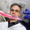
Dr. Diana Bianchi discovered that fetal cells remain in a woman's body for decades after pregnancy, perhaps indefinitely.


At the medical center, Dr. Bianchi says, her foray into the world of stem cell research involved a decade of discoveries so unexpected that despite her stellar reputation, colleagues at first looked askance.

Dr. Bianchi, who works in a lab stretched out along a narrow corridor of an old building that was once a garment factory, stumbled into the field when she was trying to find a new method of prenatal diagnosis.

She knew that a few fetal cells enter a woman's blood during pregnancy and hoped to extract those cells for prenatal diagnosis. That proved too difficult because there are so few fetal cells in maternal blood.

But then she discovered that the fetal cells do not disappear when a pregnancy ends. Instead, they remain in a woman's body for decades, perhaps indefinitely. And if a woman's tissues or organs are injured, fetal cells from her baby migrate there, divide and turn into the needed cell type, be it thyroid or liver, intestine or gallbladder, cervix or spleen.

She and her colleagues find fetal cells by looking for male cells in tissues and organs of women who have been pregnant with boys and showing that the cells' DNA matches that of the women's sons or, if the women had abortions, their male fetuses. (Cells from female fetuses also enter a woman's body, but it is quicker and easier to find the male cells by looking for cells with a Y chromosome, Dr. Bianchi says.)

One woman, for example had hepatitis C, a viral infection. But when her liver repaired itself, it used cells that were not her own.

"Her entire liver was repopulated with male cells," Dr. Bianchi said.

Such findings astonished even Dr. Bianchi. But now, with publications in leading journals, including, last month, The Journal of the American Medical Association, few doubt her.

In theory, fetal cells lurking in a woman's body are the equivalent of a new source of stem cells and could be stimulated to treat diseases. But, Dr. Bianchi says, she does not yet know for sure that the cells are stem cells - she must isolate them and prove they can turn into any of the body's specialized cells - nor where the cells reside, or how, short of injury, to spur them to action.

Using Mice and Fish, for Now

A short distance away, Dr. Leonard Zon, the chief of stem cell research at Children's Hospital, and his colleague Dr. George Q. Daley are working with stem cells from embryos, using mice and zebra fish for now. They want to learn how to transform the stem cells into immature blood cells that will divide and replenish themselves.

Then, if they can apply their work to human embryonic stem cells, they want to use the cells instead of bone marrow transplants to treat patients with genetic disorders like sickle cell anemia, and inborn disorders of the immune system.

Dr. Zon treats children with these diseases, most of whom do not have a relative whose cells match theirs closely enough to serve as a bone marrow donor. He urgently wants to help.

But he is not there yet. So far, in research that stem cell investigators say is among the most promising in the field, Dr. Zon and Dr. Daley have turned mouse embryonic stem cells into mouse blood cells. Those blood cells, however, are more mature than the ones they need, a particular type of early blood cells that can repopulate a patient's bone marrow and survive indefinitely. Ones that are more mature live out their lifespans and die within weeks.

They are also working with human embryonic stem cells, venturing into the most controversial area of stem cell work. Human embryonic stem cells are derived from human embryos, about a week old, and the only way to get the stem cells is to destroy the embryos.

Some human stem cells came from embryos that were donated by couples at fertility labs who had embryos left over after they decided their families were complete. Others came from embryos that were created to obtain stem cells; researchers paid women to donate eggs, fertilized them and let them grow to the stage where stem cells could be extracted.

The federal government has agreed to pay for research with human stem cells, but only for work with 22 lines; each line is the progeny of a single embryo. That restriction dates from Aug. 9, 2001, when President Bush issued a directive saying the government would pay for research, but only with cell lines created before that date.

Dr. James F. Battey, director of the National Institute on Deafness and Other Communication Disorders and chairman of the National Institute of Health's stem cell task force, said scientists were free to study other stem cell lines if they used private money. He understands the researchers' complaints that it would be better if the government paid for work on more lines, but, he said, as far as the federal government is concerned, "the argument isn't solely about science."

"What the president has already said on multiple occasions is that he is committed to the notion that taxpayers' money should not be used to encourage the destruction of human embryos," Dr. Battey said. "This is a White House policy."

And, he said, "it is not based solely on the needs of the scientific community."

But Dr. Zon said being able to work on more human stem cell lines could help the research.

"When you are trying to do research, you look for every advantage you can," he said. "Some embryonic stem cell lines make particular tissues better than others."

Some, for example, might more easily turn into blood cells, and others might more easily grow into nerve cells, but there is no way to know whether there is a better stem cell line for a particular cell type without trying as many as possible, Dr. Zon said. "You would want to find the line that makes the tissue you are studying."

An Embryo by Cloning

Across the river in Cambridge, in the basement of a biology building on Harvard's campus, a small group of scientists works in a two-room lab on the site of a former machine shop. Among their goals is to plunge into one of the most controversial areas of stem cell research - creating human embryos by cloning and obtaining stem cells from those embryos.

An embryo created by cloning would be an exact genetic match of the person whose cells were used to make it. Its stem cells and any mature cells derived from those stem cells would exactly match the cells in the person's body, making them perfect replacement cells.

One of the four part-time researchers at the Harvard lab, Dr. Kevin Eggan, learned to clone mice as a Ph.D. student at M.I.T. and, he said, the group is seeking approval from Harvard's ethics committee to try to start the cloning process with human cells.

The federal government forbids the use of its money to pay for such research, but this lab, directed by Dr. Douglas A. Melton, a Harvard developmental biologist, takes no federal money. Instead, the work is paid for by the Howard Hughes Foundation, the Juvenile Diabetes Foundation and the Naomi Berry Diabetes Center of Columbia University.

Cloning, however, can be onerous. In February, researchers in South Korea announced that they gotten stem cells from human embryos they created by cloning, but they began with 176 human eggs and ended up with one embryo that yielded stem cells.

Dr. Eggan, though, is not after replacement cells. His goal in cloning is to understand what goes wrong in a disease like Alzheimer's, Parkinson's or diabetes.

Dr. Melton gave an example. Suppose he had stem cells that were exact matches of 50 patients with Parkinson's disease and directed them to grow in the laboratory into nerve cells of the type that die in the disease. He could then ask when, and why, the cells die.

"Do they all show a defect at the same stage? If so, that would mean there is a common cause, like a flat tire. Or maybe each one breaks down in a different way. Is there one way to get Parkinson's, or 50 ways?" Dr. Melton asked.

"We could use that information to do drug screening," he added, possibly finding ways to prevent the nerve cell death.

For now, though, the Harvard lab is becoming a supplier to the world of its own 17 lines of human embryonic stem cells, created without cloning, and made from 286 frozen embryos created by in vitro fertilization.

Realism About Treatments

Meanwhile the national debate over the use of human embryonic stem cells goes on.

While many Americans say in polls that they favor using these cells, many others have strong moral objections. Creating and destroying a human embryo to obtain stem cells, they say, is ethically unacceptable, and doing research on human embryonic stem cell lines that are already in existence does not right the wrong.

It is "a kind of after-the-fact cooperation with this destruction," said Richard Doerflinger, deputy director of pro-life activities for the United States Conference of Catholic Bishops.

The challenge for scientists in the midst of a fierce political debate, many say, is to be realistic about how hard it is to develop treatments.

Dr. Battey lists some of the challenges ahead: getting the cells to develop into exactly the adult cells that are needed, demonstrating that the adult cells can survive, preventing rejection and controlling cell growth.

Such issues, Dr. Battey said, "need to be addressed in animal models before any thoughtful person would go into humans."

#252 kevin

  • Member, Guardian
  • 2,779 posts
  • 822

Posted 03 September 2004 - 06:21 AM

Link: http://www.eurekaler...w-bmc083104.php


Posted Image
Bone marrow cells routinely help with wound healing
'Wounds may not heal the way we thought they did'

Bone marrow produces cells that not only help fight infection, but also permanently heal wounds, according to research at the University of Washington. Previously, researchers had not known that bone marrow contributed to the development of new skin in wounds.

The findings will be published in the Sept. 3 issue of Stem Cells.

"Wounds may not heal the way we thought they did," says Dr. Richard Ikeda, a biochemist at the National Institute of General Medical Sciences, one of the National Institutes of Health, which supported the work. "This study shows that bone marrow stem cells, in addition to cells from the surrounding tissue, may actually contribute to the healing process. If this is the case, it could lead to completely new ways of treating serious wounds."

When a body is wounded, the body immediately tries to form a clot in order to stop the bleeding. The seal is formed with the help of cells that circulate in your blood all the time and are on the spot immediately. The body also has an inflammatory response: signals direct white blood cells to the area of the wound. The white blood cells arrive to fight off foreign bacteria and infection. This inflammatory response is responsible for the red area around a wound. The inflammatory response goes away within a few days to a week, assuming there is no continued infection.

"Scientists have long assumed that once the inflammatory response concludes, the white blood cells mostly either then die or go into circulation in the bloodstream. We did not know, until now, that the bone marrow-derived cells go on to become a significant part of the new skin," said Dr. Frank Isik, professor of surgery at the University of Washington. "We've known that bone marrow cells are involved in wound healing and inflammation – now we have data that shows bone marrow cells are involved in normal skin maintenance, in maintaining the matrix environment and integrity of the skin."

Bone marrow has been studied for a number of purposes in recent years because it is rich in stem cells – cells that can go on to become many different kinds of cells. In order to conduct this research, Isik and colleagues obtained a strain of mice whose bodies glow green under fluorescent light. The researchers removed bone marrow from the mice and then performed a stem cell transplant into a genetically identical strain of normal mice, whose cells do not glow green. Afterward, only the bone marrow of the transplanted mice glowed green inside the bodies of the mice, allowing researchers to track the bone marrow cells throughout the body. Researchers found green cells throughout the body, but observed that the highest concentration of bone marrow cells was in normal skin.

That was a surprise. People have known for awhile that there are a few white blood cells in the skin – that's how people come down with contact dermatitis. Contact dermatitis happens when someone develops an inflammatory reaction to a substance that touches his or her skin. However, the white blood cells involved in contact dermatitis express a certain protein, CD45. The new cells identified in the transplanted mice did not produce that protein, and do not seem to be implicated in contact dermatitis. Researchers found that even after six weeks, long after the infection-fighting role seems to be over, the bone marrow-derived cells cluster within the healing area of a wound.

The researchers ran these skin cells through a flow cytometer to separate them into green and non-green fractions and found only the green cells in the skin produced collagen type III, which is one of the two most abundant collagens in skin. The native skin cells produced only collagen type I. Researchers do not know why bone marrow would produce collagen III, which is typically found in connective tissues such as skin.

"What we have here is a new cell population that was not previously recognized," Isik said. "The bone marrow cells help form the matrix of the skin. Collagen is what gives your skin its tough nature. It's expandable, and it's tough. You cannot break your skin without a sharp object. The reason is because of the collagen content, a scaffolding that is very strong."


###
Isik is involved in follow-up studies to determine exactly what role the cells of bone marrow origin have within the skin. One question is whether diseases such as diabetes, which leads to poor wound healing, affect the function of bone marrow cells in the skin and wounds. Researchers hope that someday, scientists might use new mechanisms to promote wound healing, and troubleshoot chronic wounds.

#253 aristotle

  • Guest
  • 2 posts
  • 0

Posted 20 September 2004 - 02:32 PM

I feel that we are one step closer to regeneration of lost/diseased/dying body parts.

ANGIOGENESIS {as treatment for small vessel vascular disease}
by Debra Woodard, RN, BSN, MA
Posted 08/31/2004 in
http://www.medscape....warticle/487324

Until recently, diagnoses of Peripheral Vascular Disease (PVD) and Peripheral Arterial Disease (PAD) held a life filled with disability with a shortened life span. Now there is hope and a brighter future for the sickest of these patients as researchers are injecting failing hearts, occluded coronary arteries, and vessels in legs with a patient's own stem cells or DNA that will grow new vessels around the occlusion, thereby restoring circulation.


#254 Lazarus Long

  • Life Member, Guardian
  • 8,116 posts
  • 242
  • Location:Northern, Western Hemisphere of Earth, Usually of late, New York

Posted 29 September 2004 - 01:36 AM

Well, well, well, here comes the heavy hitters.

Dolly's Creator Applies for Human Cloning License

Tue Sep 28, 2:25 PM ET Science - Reuters
By Patricia Reaney

LONDON (Reuters) - Scientists who created Dolly the sheep, the world's first cloned mammal, applied for a license on Tuesday to clone human embryos to obtain stem cells for research into Motor Neurone Disease.

Professor Ian Wilmut, of the Roslin Institute in Edinburgh, hopes to study how the paralyzing illness -- also known as Amyotrophic Lateral Sclerosis (ALS) or Lou Gehrig's disease -- develops, with a view to finding an effective treatment.

"We believe it will produce entirely new opportunities to study Motor Neurone Disease," he told a news conference.

If the license is approved by the Human Fertilisation and Embryology Authority (HFEA), Britain's cloning watchdog, it will be the second granted for the controversial research, which has incited fierce ethical debate because it involves creating human embryos which can be mined for their stem cells.

A team of scientists from Newcastle University in northern England were granted a license in August to clone human embryos to develop new treatments for diabetes and degenerative diseases such as Alzheimer's and Parkinson's.

"This is not reproductive cloning in any way," said Professor Christopher Saw, of the Institute of Psychiatry, who will collaborate on the research.

Human reproductive cloning is outlawed in Britain but therapeutic cloning, creating embryos as a source of stem cells to cure diseases, is allowed on an approved basis. Stem cells are master cells of the body that can develop into other cell types.


DOLLY TECHNIQUE

Motor Neurone Disease affects nerve cells that carry instructions from the brain to the muscles. It weakens muscles and causes paralysis but the patient's brain is not affected.

About 70,000 patient worldwide suffer from the illness including British physicist Stephen Hawking.

The disease is inherited in about 10 percent of cases. There is no effective treatment.

Wilmut and his colleagues plan to use the same technique that was successful in creating Dolly in 1996. They will extract genetic material from a skin or blood cell of patients suffering from an inherited form of the illness and place it in an egg whose nucleus has been removed.


The egg will be stimulated to develop into an embryo and allowed to develop for about six days, when the stem cells will be extracted. The scientists will compare the stem cells with both healthy and diseased cells from patients to better understand the illness and to test potential medicines.

"Our objective is to understand the disease. We hope one day it will lead to treatments," Wilmut added.

#255 Michael

  • Advisor, Moderator
  • 1,293 posts
  • 1,792
  • Location:Location Location

Posted 13 October 2004 - 02:43 AM

All:

Hypothetical scenario: In five years the production of large quantities of ESCs and the refinement of SCNT have been developed. This allows for the transplantion of genetically identitical tissue without the risks associated with immunosupressant therapies.


I realize that this is hypothetical (you even said so :) ), but 5 years for human application is WILDLY optimistic, unfortunately. IAC:

How would this help with Type I diabetes? Wouldn't the patient still need to be placed on an immunosupressant therapy because the under lying cause of the disease lies in the faulty genetically coded instructions which causes the individual's body to attack "healthy" pancreatic islets?  Is there something I'm missing here?


A few things, from my semi-educated vantage point (more informed readers can jump in if I say anything too egregiously incorrect):

Right now, there is a transitional therapy for type I diabetes called the "Edmonton protocol" in which they transplant pancreatic cells into the patient. Yes, they have to take immunosuppressants, but it's still a real improvement in quality and quantity of life, not having to constantly shoot up, monitor glucose, occasionally go into glucose shock, suffer complications from even relatively tight (by type I
diabetes standards) blood glucose, etc etc.

Based on the successes and limitations of the Edmonton Protocol, ESC or SCNT could help improve many diabetics' lives for several reasons:

First, there is a serious lack of pancreases. ESC or SCNT could in principle eliminate this limiitation. Even if there were no solutions on either the immunity or autoimmunity front, this would be a big step forward.

Second, even if you don't in any way address the AUTOimmunity issue, you can eliminate the more immediate problem of rejection with SCNT. Right now, people taking the Edmonton protocol are having to deal with both.

Third, the above point would mean a real step forward for CHILDREN with IDDM. Presently, children just can't handle the immunosuppressants required, adn are not administered the Edmonton protocol. So for these patients, SCNT would not just IMPROVE the therapy by eliminating rejection, but would make it available for the first time.

Fourth, you can manipulate the genes of ESCs and then multiply them easily. This might allow one to genetically alter just 1 gene to prevent the expression of teh autoimmune glycoproteins in what are otherwise fully compatible pancreas cells, creating cells that WOULDN'T be attacked, and then clonally expand this cell into a pancreas whihc has neither immune nor autoimmune problems. Doing gene therapy on a whole, differentiated pancreas is both still difficult in humans, and still risky (see problems with vectors causing cancer in bubble babies).

Finally, with many cell types -- neurons and myocytes especially -- the differentiated cells don't culture well and the model is not physiologic. Having cloned stem cells allws one to propagate them and study them in culture, letting us learn more about the disease's pathogenesis even if we never use the cells directly for regenerative medicine.

Unless, of course, the technology was proficient enough to constantly rejuvinate a patient's continually depleting beta cells...


If worse came to worst, in principle, one could do just that.

-Michael

#256 DJS

  • Guest
  • 5,798 posts
  • 11
  • Location:Taipei
  • NO

Posted 13 October 2004 - 10:39 PM

Michael,

Thank you! Your response has definitely enhanced my understanding of stem cell. :) Let's see...yes, I am aware that 5 years is overly optimistic -- I should have said 10 years (and yes I know, that would still be considered overly optimistic by some! [lol] ). I am also aware of the Edmonton Protocol and have seen it referenced several times in the literature I have read on diabetes, but after reading your post I think I will commit to more research on the subject (if I can ever find the time [:o] ).

This segment of your post was particularly valuable to me:

Third, the above point would mean a real step forward for CHILDREN with IDDM.  Presently children just can't handle the immunosuppressants required...


You make a valid point, and one that I had failed to consider up until now. So point taken [thumb]

One question I would be interested in seeing your answer to (just for shits and giggles) is what a *realistic* time table would be for real break throughs in stem cell technology.

Nice to make your acquaintance,

DonS

#257 Cyto

  • Topic Starter
  • Guest
  • 1,096 posts
  • 1

Posted 30 October 2004 - 09:07 PM

Human embryonic stem cells promising for replacement of blood supply

Posted Image

Researchers at the University of Minnesota Stem Cell Institute are one step closer to understanding how blood cells develop through the use of human embryonic stem cells. The research better defines the conditions under which blood cell development occurs, making the process easier to replicate. The findings are published in the October issue of Experimental Hematology.

"These findings do more than give us a basic understanding of blood cell replacement--they allow us to consider potential future therapies," said Dan Kaufman, M.D., assistant professor of medicine in the division of hematology, oncology and lead researcher. "We can envision blood therapies completely compatible with the patient, such as use of embryonic stem cells to make red blood cells for platelets used in blood transfusions, or a source of new blood supply free of any viruses. They might also be a source for bone marrow transplants, especially for those patients who do not otherwise have an appropriately matched donor."

This process is also significant because the blood cells were developed without the use of animal serum, which was previously thought to be essential for blood cell development. Instead, specific growth factors are added to guide the cell differentiation. These results are important for potential human application. Animal serum can potentially contaminate findings and create complications for human trials.

Kaufman's research interests focus on hematopoietic and endothelial cell development from human and non-human primate embryonic stem cells. This research uses embryonic stem cells to understand the earliest stages of blood cell development.

#258 Cyto

  • Topic Starter
  • Guest
  • 1,096 posts
  • 1

Posted 01 November 2004 - 06:24 AM

Sick kids researchers unmask the potential of stem cells found in adult skin

Posted Image

Researchers at The Hospital for Sick Children (Sick Kids) have shown that stem cells found in adult skin retain their embryonic capability of making many types of cells. This discovery affirms the potential that stem cells derived from this non-controversial source possess for the development of possible therapies for spinal cord injury and nervous system disorders. This research is reported in the scientific journal Nature Cell Biology, published online October 31, 2004.

"We think these stem cells are actually embryonic cells that go out into the skin during development and then stay in reservoirs in hair follicles," said Dr. Freda Miller, the study's principal investigator, a senior scientist in Development Biology in the Sick Kids Research Institute and a professor of Molecular and Medical Genetics, and Physiology at the University of Toronto.

"These stem cells are similar to a type of embryonic stem cell called a neural crest stem cell, and like neural crest stem cells, are endogenous and multipotent in nature. These neural crest stem cells generate the peripheral nervous system, and we are therefore now confident that we can make neural and other types of cells from the stem cells found in adult skin," added Dr. Miller, also Canada Research Chair in Developmental Neurobiology.

The research team can now predict what type of cells can be made from these stem cells (called skin-derived precursors, or SKPs) based on the role played by neural-crest stem cells during embryogenesis. In addition to generating the peripheral nervous system, neural crest stem cells generate other tissues such as bone, cartilage, some types of muscle, and even part of the heart. This research was conducted in mice, with similar findings made recently by Dr. Miller's group in the human cells.

"The cells that Dr. Miller's group has found in the skin have huge potential to treat brain disorders because they are capable of transforming into neurons normally only found in the brain and other nervous tissue. This new research provides an explanation for the cells' ability to make neurons and further enhances our understanding of a potentially valuable cell type for stem cell therapy," said Dr. Ron Worton, scientific director of Canada's Stem Cell Network. "The Stem Cell Network is pleased to have supported this work in Dr. Miller's laboratory."

#259 Cyto

  • Topic Starter
  • Guest
  • 1,096 posts
  • 1

Posted 07 November 2004 - 04:56 PM

Stem Cell Researcher to receive research award from Biomedical Group

Treena Livingston Arinzeh, PhD, an assistant professor of biomedical engineering at New Jersey Institute of Technology (NJIT) whose research has proven the potential of adult stem cell research to help patients suffering from spinal cord injuries and related diseases, will receive an Outstanding Women in Research Award from The New Jersey Association for Biomedical Research (NJABR), Union.

Arinzeh, 34, is one of the leading stem cell researchers in the nation. In October, she was awarded the nation's highest honor given to young scientists and engineers by President Bush ([pirate]). Adult stem cells have miraculous potential, and three years ago, Arinzeh published a paper in the Journal of Biomedical Materials Research that documented a breakthrough in stem cell research. The paper focused on developing scaffolds that aid stem cells. Scaffolds are biomaterials, such as calcium phosphates, that act as frameworks for stem cells, allowing the cells to repair bone.

Arinzeh performed animal studies on rats with bone defects; she also performed cell-culture studies. After 12 weeks, their bones were regenerated, with full restoration of the mechanical properties of their long bones. Her studies could lead to medical breakthroughs that would help a host of patients. Stem cell implantation, for instance, could help cancer patients who've had large tumors removed from bone, Arinzeh says. Stem cells could also help patients suffering from osteoporosis.

Perhaps most importantly, Arinzeh published another paper in the Journal of Bone and Joint Surgery proving that adult stem cells taken from one patient can be successfully implanted in another. Researchers originally thought such a transfer might be rejected. And it's not just defective bones that may be regenerated by stem cells and biomaterials. Arinzeh is now testing biomaterials that, in combination with adult stem cells, might also repair cartilage, tendon and neuronal tissues.

"This is a very exciting time to be doing stem cell research," Arinzeh said. "The field is wide open and has the potential to influence how physicians treat patients with severely damaged or diseased tissues."

#260 Cyto

  • Topic Starter
  • Guest
  • 1,096 posts
  • 1

Posted 17 November 2004 - 10:54 PM

Cancer Stem Cells Initiate And Grow Brain Tumours

"Now that we have confirmed that a small number of cancer stem cells initiates and maintains human brain tumour growth in a mouse model, we can potentially use the mouse model with each patient's tumour cells to see if therapies are working to conquer that patient's tumour," said Dr. Peter Dirks, the study's principal investigator, a scientist and neurosurgeon at Sick Kids, and an assistant professor of Neurosurgery at U of T. "A functional analysis of the brain tumour stem cell may also give new insight into patient prognosis that may then warrant individual tailoring of therapy."

Dr. Dirks' laboratory was able to regrow an exact replica of patients' brain tumours in a mouse from the isolated cancer stem cells, or brain tumour initiating cells. They were then able to study the growth of the human brain tumour in the mouse model using the advanced imaging technology in the Mouse Imaging Centre (MiCE) at Sick Kids.

Brain tumours are the leading cause of cancer mortality in children and remain difficult to cure despite advances in surgery and drug treatments. In adults, most brain tumours are also among the harshest cancers with formidable resistance to most therapies.

"Next, we are going to study the gene expression of the brain tumour stem cells. Once we have identified what genes are expressed in those cells, we will then be able to target these genes using new drugs or genetic-type therapies," said Dr. Sheila Singh, the paper's lead author and Sick Kids neurosurgery resident and U of T graduate student who is enrolled in Sick Kids' Clinician-Scientist Training Program. Dr. Singh was supported by a fellowship from The Terry Fox Foundation, as well as by funding from the Neurosurgical Research and Education Foundation and the American Brain Tumor Association.

"We have shown that it is really worthwhile to invest further in studying brain tumour stem cells, as we will be able to determine if current therapies are failing because they are not stopping the cancer stem cells," added Dr. Dirks. "It also looks like cancer stem cells play a role in other solid tumours such as breast cancer, so we can all work together to develop new treatments for these cancers."

#261 Cyto

  • Topic Starter
  • Guest
  • 1,096 posts
  • 1

Posted 22 November 2004 - 09:12 PM

UCI researchers use human embryonic stem cells to create new nerve insulation tissue that can aid spinal cord repair

Irvine, Calif, November 22, 2004

For the first time, researchers have used human embryonic stem cells to create new insulating tissue for nerve fibers in a live animal model – a finding that has potentially important implications for treatment of spinal cord injury and multiple sclerosis.

Researchers at the UC Irvine Reeve-Irvine Research Center used human embryonic stem cells to create cells called oligodendrocytes, which are the building blocks of the myelin tissue that wraps around and insulates nerve fibers. This tissue is critical for maintenance of proper nerve signaling in the central nervous system, and, when it is stripped away through injury or disease, sensory and motor deficiencies and, in some cases, paralysis result.

In this study, neurologist Hans Keirstead and colleagues at UCI and the Geron Corporation ( [:o] Their still alive!) devised a novel technique that allows human embryonic stem cells to differentiate into high-purity, early-stage oligodendrocyte cells. The researchers then injected these cells into the spinal cords of mice genetically engineered to have no myelin tissue.

After transplantation into mice, the early-stage cells formed into full-grown oligodendrocyte cells and migrated to appropriate neuronal sites within the spinal cord. More importantly, the researchers discovered the oligodendrocyte cells forming patches of myelin’s basic protein, and they observed compact myelin tissue wrapping around neurons in the spinal cord. These studies demonstrated that the oligodendrocytes derived from human embryonic stem cells can function in a living system.

Results of this study are published online in the peer-reviewed journal Glia.

“These results are extremely exciting and show great promise,” Keirstead said. “What we plan to do next is see how these cells improve sensory and motor function, and hopefully it will lead to further tests with people who suffer from these debilitating illnesses and injuries.”

Gabriel I. Nistor and Minodora O. Totoiu from UCI collaborated with Nadia Haque and Melissa K. Carpenter of the Geron Corporation on the study, which was supported by Geron, UC Discovery, Research for Cure and the Reeve-Irvine Research Center. Geron provided the human embryonic stem cells used in this study.

In previous studies, Keirstead and colleagues have identified how the body’s immune system attacks and destroys myelin tissue during spinal cord injury or disease states. They’ve also shown that, when treated with antibodies to block immune system response, myelin is capable of regenerating, which ultimately restores sensory and motor activity.

The Reeve-Irvine Research Center was established to study how injuries and diseases traumatize the spinal cord and result in paralysis or other loss of neurologic function, with the goal of finding cures. It also facilitates the coordination and cooperation of scientists around the world seeking cures for paraplegia, quadriplegia and other diseases impacting neurological function. Named in honor of Christopher Reeve, the center is part of the UCI College of Medicine.

#262 Cyto

  • Topic Starter
  • Guest
  • 1,096 posts
  • 1

Posted 29 November 2004 - 10:58 PM

Quality control my friends, quality control. :)


Finding Could Improve Safety of Stem Cell Transplants

Posted Image

A lipid that helps destroy potentially harmful cells during brain development shows promise for improving the safety and efficacy of stem cell transplants, say researchers at the Medical College of Georgia and University of Georgia.

When embryonic stem cells are being coaxed toward becoming brain cells that could be transplanted, that lipid, ceramide, helps eliminate cells that could later form tumors called teratomas, researchers say in the Nov. 22 issue of The Journal of Cell Biology.

“The body has amazing mechanisms to eliminate cells that are no longer wanted and that if they remain will harm the body by developing into tissues that are not meant to be,” says Dr. Erhard Bieberich, MCG biochemist and the study’s lead author. “Our studies show this particular mechanism can help stem cells safely become the cells we want them to be.”

“This is another approach to controlling differentiation and getting the cell types that you want,” says Dr. Brian G. Condie, developmental neurobiologist at UGA and MCG and senior author on the paper.

While it’s the ability of embryonic stem cells to make all types of tissue -- from brain cells to heart cells -- that has scientists worldwide exploring their potential to treat devastating diseases, their pluripotency can also be harmful if uncontrolled, says Dr. Bieberich.

Drs. Bieberich and Condie demonstrated in the Aug. 4, 2003 issue of The Journal of Cell Biology that a natural process occurs during development to eliminate excessive and potentially harmful cells. Just before neurons begin forming, there is a massive production of proteins and up-regulation of lipids. At that point, about half the cells have high levels of the protein PAR-4, half have high levels of the protein, nestin, and all have high levels of ceramide.

The researchers found cells that inherited PAR-4 died when partnered with ceramide. Fortunately, the nestin-bearing cells are most likely to become neurons while the PAR-4 cells, should they survive, could contribute to brain malformation.

**In this new paper, they took their findings in mouse embryonic stem cells and also looked at an approved line of human embryonic stem cells available through the National Institutes of Health Embryonic Stem Cell Registry.**

Posted Image
Neuroprogenitor cells in culture that will become neurons.

Posted Image
Cells in culture with neurons stained green, supporting glial cells stained red and cell nuclei stained blue.

They found as the cells differentiated in culture, those containing PAR-4 have yet another bad dance card.

“We have discovered that particular cells derived from embryonic stem cells that express PAR-4 also cause teratoma formation,” says Dr. Bieberich of the mostly benign growths comprised of multiple types of tissue, typically none of which belong in the tissue where they are found.

They found PAR-4-expressing cells also express Oct-4, a transcription factor that controls a cell’s ability to develop into all three basic types of tissue: mesoderm, ectoderm and endoderm. “If Oct-4 is expressed, the cells are still pluripotent, which is good if you want to grow all those kinds of embryonic layers,” says Dr. Bieberich. “But if you transplant them, you are at risk of forming teratomas.”

However, at least in the culture dish, when they added PAR-4’s lethal dance partner, ceramide, to the mix, PAR-4- and Oct-4-expressing cells again died before they could do harm.

The ceramide analogue, N-oleoyl serinol, or S18, also increased the proportion of nestin-containing cells in cell cultures and grafts.

Drs. Bieberich and Condie were quick to note that in their studies, they intentionally left PAR-4- and Oct-4-bearing cells in the mix to see if they could eliminate them.

“There already are ways to grow stem cells, purify them in cell culture and get a pure population of stem cells that you can transplant,” says Dr. Condie. “You want to make those cells differentiate into a particular cell type that is no longer able to form teratomas,” Dr. Bieberich says of this purification. “Having said that, that may not always be absolute.”

“What we are trying to do is find ways that can be combined with those methods currently being used to further reduce the chances of teratoma formation and make stem cells extremely safe,” Dr. Condie says. “This is something that you want to have zero doubt about.”

The next step is to look at an intact mouse embryo to see if the identical processes are at work.

Link

#263 Cyto

  • Topic Starter
  • Guest
  • 1,096 posts
  • 1

Posted 09 December 2004 - 07:52 PM

Neural crest stem cells in skin could provide alternative to embryonic stem cell use

Posted Image
(FYI: It points out where the crest is, otherwise it has nothing to do with this news)


Cell replacement therapy offers a novel and powerful medical technology. A type of embryonic stem cell, called a neural crest stem cell, that persists into adulthood in hair follicles was recently discovered by Maya Sieber-Blum, Ph.D., of the Medical College of Wisconsin, Milos Grim, MD Ph.D., of Charles University Prague, and their collaborators.

The discovery – reported recently in Developmental Dynamics, a journal of the American Association of Anatomists published by John Wiley & Sons, Inc. – may in many instances provide a non-controversial substitute for embryonic stem cells. Embryonic stem cells are unique, because they can differentiate into any cell type of the body. Their use, however, raises ethical concerns because embryos are being destroyed in the process. In contrast, neural crest stem cells from adults have several advantages: similar to embryonic stem cells, they have the innate ability to differentiate into many diverse cell types; they are easily accessible in the skin of adults; and the patient's own neural crest stem cells could be used for cell therapy. The latter avoids both rejection of the implant and graft-versus-host disease.

Studies in the mouse showed that neural crest stem cells from adult hair follicles are able to differentiate into neurons, nerve supporting cells, cartilage/bone cells, smooth muscle cells, and pigment cells. Preliminary data indicate that equivalent stem cells reside in human hair follicles.

"The goal of our research is to apply neural crest stem cells from adult hair follicles in cell replacement therapy in selected instances," Sieber-Blum says. This may include, spinal cord injury, Parkinson's disease, multiple sclerosis, Hirschsprung's disease, peripheral neuropathies, certain defects of the heart, and bone degeneration. Though promising, this research is still in the animal testing stage. Additional research is required before it could benefit patients.


Link

#264 manofsan

  • Guest
  • 1,223 posts
  • 56

Posted 18 December 2004 - 01:10 PM

Stem cells used to rebuild girl's skull:

http://www.cnn.com/2...s.ap/index.html

Another example of stem cells taking their cues on what to differentiate into from local differentiated cells.

#265 manofsan

  • Guest
  • 1,223 posts
  • 56

Posted 18 December 2004 - 02:45 PM

Stem cells repairing bad livers:

http://www.newscient...le.ns?id=dn6804

Hey, why stop there? Why not fix bad kidneys, pancreas -- gee, the sky's the limit. ;)

#266 Cyto

  • Topic Starter
  • Guest
  • 1,096 posts
  • 1

Posted 24 January 2005 - 03:55 AM

Current human embryonic stem cell lines contaminated UCSD/Salk team finds

Currently available lines of human embryonic stem cells have been contaminated with a non-human molecule that compromises their potential therapeutic use in human subjects, according to research by investigators at the University of California, San Diego (UCSD) School of Medicine and the Salk Institute in La Jolla, California.

In a study published online January 23, 2005 in the journal Nature Medicine, the researchers found that human embryonic stem cells, including those currently approved for study under federal funding in the U.S., contain a non-human, cell-surface sialic acid called N-glycolylneuraminic acid (Neu5Gc), even though human cells are genetically unable to make it. In a related paper published November 29, 2004 by the Journal of Biological Chemistry (JBC), the Varki group has also discovered the exact cellular mechanism by which this occurs.

In studies with one of the federally approved human embryonic stem cell lines, the investigators determined that the Neu5Gc is incorporated by the stem cells when they are grown or derived from laboratory cultures that contain animal sources of the non-human Neu5Gc molecule. All traditional culture-dish methods used to grow all human embryonic stem cells include animal-derived materials, including connective tissue cells (so-called "feeder layers") from mice and fetal calf serum.

"The human embryonic stem cells remained contaminated by Neu5Gc even when grown in special culture conditions with commercially available serum replacements, apparently because these are also derived from animal products," said both papers' senior author Ajit Varki, M.D., UCSD professor of medicine and cellular & molecular medicine, and co-director of the UCSD Glycobiology Research and Training Center.

The research in Nature Medicine was done with human embryonic stem cells grown in the laboratory of Fred Gage, Ph.D., professor, Laboratory of Genetics, the Salk Institute, La Jolla, California, an adjunct professor of neurosciences at UCSD, and an author on the Nature Medicine paper.

Previously, the Varki lab found in 1998 that humans are uniquely different from other mammals studied in that people do not express Neu5Gc*. In a 2003 study**, the UCSD researchers found that humans have naturally occurring antibodies that are directed against Neu5Gc. In the current Nature Medicine paper, the scientists found that the human embryonic stem cells contaminated with Neu5Gc became, effectively, like animal cells, being attacked by human antibodies, and thus rendering them useless as a potential therapeutic tool in humans.

"It's an important safety issue because this opens up the idea that metabolic transfer of glycans is occurring between cells," said Gage. "Also, components of the growth medium have the capacity to change the immunological characteristics of the human ES cells. More research is needed to understand the optimal conditions for preparing human cells for therapeutic application."

"We considered that one partial solution to the problem was to use human serum in the growth medium," Varki said. When the team grew the cells in heat-inactivated human serum specially selected for low concentrations of anti-Neu5Gc antibodies, the immune response was significantly reduced, but not completely eliminated.

In their experiments, the researchers used recently developed probes to detect the presence of Neu5Gc on the cell surface of human embryonic stem cells that had been grown in traditional culture conditions. The scientists further confirmed the presence of Neu5Gc with a process called electrospray mass spectrometry. The percentage of total sialic acids present as Neu5Gc in the embryonic stem cells varied from 2.5 to 10.5 percent. In human embryonic stem cells that had been allowed to differentiate into embryoid bodies (EB), which is the first step in preparing them for potential use in humans, the percentage of total sialic acids present still ranged from 5 to 17 percent.

Varki and his team noted that many efforts have been made during the last few years to try to eliminate any animal-derived culture components in human stem cell culture. However, many of the specialized media used for growth and differentiation still contain materials from animal sources and are hence contaminated with Neu5Gc.

In addition to using human serum, the researchers suggested the possibility of using what are called "feeder cells" from mice with a human-like defect in Neu5Gc production. They noted that they have recently produced such a mouse. Another possibility being attempted by groups in other parts of the world is to use human embryo-derived connective tissue cells as the feeder layer in the culture.

A further solution might be a short-term culture in heat-inactivated serum from the actual patient who is going to receive the therapy, the scientists said. However, it may still prove difficult to completely eliminate the Neu5Gc, because is has become metabolically incorporated into the currently available, federally-funded human embryonic stem cell lines.

"With this discovery, the preexisting general concern about using animal products for deriving human embryonic stem cells has become more specific, being defined in molecular terms," Varki said.

"Such issues will, of course, become irrelevant if complete elimination of Neu5Gc can be achieved by deriving new human embryonic stem cells that have never been exposed to Neu5Gc-containing animal products of any kind," the researchers said in the Nature Medicine paper, noting that none of the suggested approaches guarantees the complete elimination of Neu5Gc from existing cultures. "Therefore, it would seem best to start over again with newly derived human embryonic stem cells that have never been exposed to any animal products, and ideally, only ever exposed to serum from the intended transplant recipient."

"However, such an approach could not be pursued under existing rules for the use of federal grant dollars," Varki said.

The first author of the Nature Medicine study is Maria J. Martin, Ph.D., a post doctoral researcher in Varki's lab at UCSD. An important additional author is Gage's post doctoral fellow Alysson Muotri, Ph.D. The study was funded by the National Institute of General Medical Sciences at the National Institutes of Health, the Lookout Fund, and by the G. Harold and Leila Y. Mathers Charitable Foundation of New York.

In addition to Varki, authors of the related study in JBC included Muriel Bardor, Ph.D. and Dzung Nguyen, Ph.D., post-doctoral fellows, and Sandra Diaz, a research associate. They determined that Neu5Gc gets into human cells by being engulfed in fluid droplets and then moved to the cytoplasm of the cell by a "pump" called the lysosomal sialic acid transporter.

Varki noted that this pathway is an unusual and previously unknown one that may also be relevant to the entry of other small molecules into cells. In addition, the JBC study showed how Neu5Gc attached to dietary proteins from animals could be incorporated into cells lining the stomach and colon, organs where consumption of red meat has been associated with risk of cancer.

"Knowing the mechanism that this molecule uses to get into human cells may give us clues to possible solutions to the problems that it may cause in various situations," Varki said.

#267 Cyto

  • Topic Starter
  • Guest
  • 1,096 posts
  • 1

Posted 24 January 2005 - 09:21 PM

PRIMING EMBRYONIC STEM CELLS TO FULFILL THEIR PROMISE

Posted Image

Bioengineering researchers at the University of California, San Diego have invented a process to help turn embryonic stem cells into the types of specialized cells being sought as possible treatments for dozens of human diseases and health conditions. Sangeeta Bhatia and Shu Chien, UCSD bioengineering professors, and Christopher J. Flaim, a bioengineering graduate student, described the cell-culture technique in a paper published in the February issue of Nature Methods, which became available online on Jan. 21.

Embryonic stem cells are considered the blank-slate, raw material needed to repair or replace damaged or missing liver, nerve, muscle, and other tissues and organs. However, in order to fulfill their therapeutic promise, scientists believe that stem cells must first be coaxed to differentiate, or mature, into precursors of specialized cell types found in the body.

Embryonic stem cell differentiation is complex and far from fully understood. Scientists are focusing on four types of external inputs known to be involved in triggering the differentiation of stem cells: soluble growth factors, adjacent cells, mechanical forces, and extracellular matrix proteins that form the support structure of almost all tissues. Bhatia, Chien, and Flaim focused on just one — the extracellular matrix.

“We kept the other factors constant and developed a miniaturized technique to precisely vary extracellular matrix proteins as a way to identify which combinations were optimal in producing differentiated cells from stem cells,” said Bhatia. She, Chien, and Flaim described in their paper a technique that enabled them to identify the precise mix of proteins that optimally prompted mouse embryonic stem cells to begin the differentiation process into liver cells. Bhatia, Chien, and Flaim designed the technique with other cell biologists in mind so that any of them could duplicate it with off-the-shelf chemicals and standardized laboratory machinery. “We think other researchers could easily use this technique with any other tissue in mouse, or human, or any other species,” said Bhatia.

Scientists have identified about 100 proteins — including laminin, fibronectin, and several kinds of collagen — that function as the extracellular matrix, or scaffolding, of most mammalian tissues. Until now, there has been no practical way to evaluate the daunting number of possible combinations of these proteins, any one of which could form the optimal “niche” for a desired type of differentiated cell.

In their experiments, the UCSD researchers took advantage of the knowledge that the extracellular matrix in liver is comprised primarily of just five proteins. They applied spots of all 32 possible combinations of the five proteins as engineered niches onto the surface of gel-coated slides, and then added mouse embryonic stem cells to the niches. After the cells were allowed to grow, the researchers assayed their progression into liver cells. “We looked at all the combinations at once,” said Bhatia. “Nobody has done this combinatorial approach before.”

Bhatia, Chien, and Flaim reported that either collagen-1 or fibronectin had strongly positive effects on the differentiation of the stem cells they tested. Unexpectedly however, when both collagen-1 and fibronectin were combined in one niche, the liver cell differentiation rate was subtly inhibited. “You would not predict that from the customary cell biology experiments,” said Bhatia. “By using this combinatorial technique we were surprised to find many interesting interactions, and we were able to tease out the effects of each protein, alone and in combination with others.”

Cell biologists have not performed such combinatorial assays for other desired cell types because they have no practical way to do so. Bhatia, Chien, and Flaim seized on the unique ability of so-called DNA spotting machines to deliver tiny volumes of liquid, about one trillionth of a liter per spot. The spotting machines, which cost as little as $20,000, have become common fixtures at most research universities, but the innovation reported today in Nature Methods involved using such a machine to spot solutions of proteins rather than DNA. The UCSD researchers also refined other parameters so that the technique would be reproducible in other research laboratories.

“When we talked to our colleagues, it was clear that, whether it’s cells in the liver, brain, or heart, there had been no practical way for researchers to find the optimal extracellular matrix needed to turn embryonic stem cells into cells with therapeutic potential,” said Bhatia. “We think we’ve developed an enabling technology for stem cell research and other areas of cell biology in the sense that all of a sudden scientists can use inexpensive and widely available reagents and machinery to optimize the conditions needed to optimize embryonic stem cell differentiation.”

Bhatia is planning further studies on generating liver cells from embryonic stem cells to make an artificial liver. She plans to seek funding to further her artificial liver research from the new California Institute for Regenerative Medicine. The institute was created after California voters in November approved Proposition 71, a measure that authorized the state to borrow $3 billion to fund stem cell research over the next 10 years.

“I’m really excited about the stem cell applications of our new technology,” said Bhatia. “We feel that this extracellular matrix part of the stem cell niche has been understudied. If we can now take what we’ve learned, add combinations of growth factors, and even add other cells to embryonic stem cells, we may be able for the first time to study all the dimensions of the niches required to very specifically control embryonic stem cell differentiation.”

#268 Chip

  • Guest
  • 387 posts
  • 0

Posted 25 January 2005 - 12:04 AM

Well, at least maybe they can tap the California funds for stem cell research cause the federal funds are for cells that appear to be contaminated. Still, appears that mainly commercial interests are controlling the disbursement of California funds. Oh well, looks like I better load that Mandarin Chinese language teaching program and get on the ball...

http://www.businessw..._7430_tc024.htm

#269 Matt

  • Guest
  • 2,862 posts
  • 149
  • Location:United Kingdom
  • NO

Posted 25 January 2005 - 02:54 AM

I live in the UK where stem cell research is funded and not as many restrictions as the U.S

Has bush limited funds of stem cell research because of his religious beliefs? I dont understand why America is really against this.

Is america going to be left behind on this technology and have to play catch up after bush goes...?

Click HERE to rent this BIOSCIENCE adspot to support LongeCity (this will replace the google ad above).

#270 Cyto

  • Topic Starter
  • Guest
  • 1,096 posts
  • 1

Posted 25 January 2005 - 03:16 AM

I hear ya Chip, I hear ya. How much was that per year again, of the 3 billion? [huh]

and whoa182:

Bush is a faith-based man who thinks that somatic cell nuclear transfer (you may see it as the easier to type SCNT) is killing a human, or potential human. This posistion has one side saying "potentialy it can be a human" and the other side "its not going to turn into one in the petri dish." And if you look at the thread on Therapeutic Cloning, Does it violate human dignity? you will see it can be messy, I have yet to read the thread though [wis]. So we see the faith-based initiatives setting in and thus restricting a very interesting science that allows us to work with something that can regenerate atropic perturbations in a system. Adult stem cell research is still going fine since it has no restrictions, that I know of, and we will have to play "catch-up." Course I don't know who is going to be sharing information...this restriction made quite a bit of ESC researchers move elsewhere...they could keep in touch with friends here as well. (shrug) We will see, and I worry about the next Presidential elections...worry a lot.

And people don't skewer my quotes from the sides, I know there are other ways of phrasing both.


1 user(s) are reading this topic

0 members, 1 guests, 0 anonymous users