• Log in with Facebook Log in with Twitter Log In with Google      Sign In    
  • Create Account
  LongeCity
              Advocacy & Research for Unlimited Lifespans

Photo
* * * * * 1 votes

Increasing the expression of NGF/BDNF in the PRE-FRONTAL cortex?

ngf bdnf pre-frontal cortex expression

  • Please log in to reply
10 replies to this topic

#1 Rior

  • Guest
  • 279 posts
  • 71
  • Location:Interwebs

Posted 28 September 2012 - 11:13 PM


I've seen many people referencing different drugs and nootropics that seem to increase the expression of BDNF and NGF in primarily the hippocampus, (highly implicated in the formation of memory) however I haven't seen much talk of stimulating NGF and BDNF in the pre-frontal cortex. Going off of basic logic, I'd think that increasing the expression of neurotrophic factors in the hippocampus would substantially help memory, however it wouldn't have a very drastic effect on creativity/logical ability. However, if one were to focus on increasing neurotrophic factors in the pre-frontal cortex, would that not theoretically enhance creativity and logical thinking?

I did a quick search to see if I could find anything that increases BDNF/NGF in the PFC, and so far have only found that Cymbalta (SNRI) seems to cause an increase in the PFC. I would think it safe to then say that other SNRIs would cause a similar effect, however I'd rather find something that isn't an SNRI to do so.

Here's a study regarding Cymbalta:

Cell Mol Neurobiol. 2008 May;28(3):457-68. Epub 2008 Jan 3.
BDNF level in the rat prefrontal cortex increases following chronic but not acute treatment with duloxetine, a dual acting inhibitor of noradrenaline and serotonin re-uptake.

Mannari C, Origlia N, Scatena A, Del Debbio A, Catena M, Dell'agnello G, Barraco A, Giovannini L, Dell'osso L, Domenici L, Piccinni A.


Source

Department of Neuroscience, Sect. Pharmacology, University of Pisa, Pisa, Italy.


Abstract

AIMS:

Brain-Derived Neurotrophic Factor (BDNF) has a central role in neuronal survival, differentiation, and plasticity. The brain level of BDNF is changed by several mood stabilizers and antidepressant drugs acting on neurotransmitters such as noradrenaline and serotonin. We investigated the effects of acute and chronic treatment with Duloxetine, a new drug blocking the re-uptake of serotonin and noradrenaline (SNRI), on BDNF level in the prefrontal cortex, cerebrospinal fluid, plasma, and serum.
METHODS:

Wistar male rats were treated with acute (single treatment) and chronic oral administration (14 days) of different concentrations of Duloxetine (10, 30, and 100 mg/kg/day). At the end of the treatment periods, samples of blood, CSF and the prefrontal cortex were collected. BDNF levels were measured by ELISA. Levels of mature and precursor form of BDNF were measured by Western blot analysis.
RESULTS:

Animals treated with the Duloxetine at all concentrations and examined after 1 and 24 h (single treatment) did not reveal a significant change in the total BDNF level. In animals treated for 14 days with Duloxetine at 30 and 100 mg/kg, the total BDNF level increased significantly in the prefrontal cortex and CSF, but not in the plasma and serum. Using a specific antibody and Western blot we showed that the mature, but not the precursor, form of BDNF was significantly increased in the prefrontal cortex of rats treated for 14 days with Duloxetine at 30 mg/kg/day.
CONCLUSIONS:

Our results show a major finding that repeated, but not single, Duloxetine treatment increases the level of BDNF in the prefrontal cortex.


PMID: 18172756 [PubMed - indexed for MEDLINE]


#2 noot_in_the_sky

  • Guest
  • 287 posts
  • 17
  • Location:Mex

Posted 22 December 2012 - 03:24 PM

Very interesting finding Rior. :cool:

In this thread: http://www.longecity...timulates-bdnf/

Computethis suggest using ALCAR to increase Acetyl-CoA, which is responsible for the conversion of serotonin into N-acetylserotonin. So perhaps by compining both Duloxetine and ALCAR we shall get a synergetic effect.

As for the none SRI, I found this reports:

https://en.wikibooks...tic_Stimulation

Allegations that ECT may cause brain damage have been consistently refuted. In fact, it seems that ECT may stimulate an increased production of neurotrophic growth factors such as brain derived neurotrophic factor (BDNF) causing migration and proliferation of progenitor cells and growth of new neurons in the hippocampus. These findings are consistent with evidence of similar effects of various other antidepressant treatments and may be the final common pathway of the antidepressant effects.


https://www.ncbi.nlm...pubmed/23201339

Magnetic stimulation modulates structural synaptic plasticity and regulates BDNF-TrkB signal pathway in cultured hippocampal neuron

Ma J, Zhang Z, Su Y, Kang L, Geng D, Wang Y, Luan F, Wang M, Cui H.

Source

Deparment of Anatomy, Hebei Medical University, Shijiazhuang 050017, Hebei, PR China; Hebei Key Laboratory for Brain Aging and Cognitive Neuroscience, The First Hospital of Hebei Medical University, Shijiazhuang 050031, Hebei, PR China; Hebei Institute of Physical Education, Shijiazhuang 050041, Hebei, PR China.

Abstract

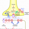
Repetitive transcranial magnetic stimulation (rTMS) is a neuropsychiatric tool that can be used to investigate the neurobiology of learning and cognitive function. Few studies have examined the effects of low frequency (⩽1Hz) magnetic stimulation (MS) on structural synaptic plasticity of neurons in vitro, thus, the current study examined its effects on hippocampal neuron and synapse morphology, as well as synaptic protein markers and signaling pathways. Similarly, both intensities of low frequency magnetic stimulation (1Hz) activated brain-derived neurotrophic factor (BDNF) and tropomyosin-related kinase B (TrkB) pathways, including the pathways for mitogen-activated protein kinase (MAPK)/extracellular signal-regulated kinase (ERK) and for phosphoinositide 3-kinase (PI3K)/protein kinase B (Akt). Specifically, low intensity magnetic stimulation (LIMS, 1.14Tesla, 1Hz) promoted more extensive dendritic and axonal arborization, as well as increasing synapses density, thickening PSD (post synaptic density) and upregulation of synaptophysin (SYN), growth associated protein 43 (GAP43) and post synaptic density 95 (PSD95). Conversely, high intensity magnetic stimulation (HIMS, 1.55Tesla, 1Hz) appeared to be detrimental, reducing dendritic and axonal arborization and causing apparent structural damage, including thinning of PSD, less synapses and disordered synaptic structure, as well as upregulation of GAP43 and PSD95, possibly for their ability to mitigate dysfunction. In conclusion, we infers that low frequency magnetic stimulation participates in regulating structural synaptic plasticity of hippocampal neurons via the activation of BDNF-TrkB signaling pathways.
Copyright © 2012. Published by Elsevier Ltd.


https://www.ncbi.nlm...pubmed/21795553

Repetitive transcranial magnetic stimulation enhances BDNF-TrkB signaling in both brain and lymphocyte.

Wang HY, Crupi D, Liu J, Stucky A, Cruciata G, Di Rocco A, Friedman E, Quartarone A, Ghilardi MF.

Source

Department of Physiology, Pharmacology & Neuroscience, Sophie Davis School of Biomedical Education, The City University of New York Medical School, New York, New York 10031, USA. hywang@sci.ccny.cuny.edu

Abstract

Repetitive transcranial magnetic stimulation (rTMS) induces neuronal long-term potentiation or depression. Although brain-derived neurotrophic factor (BDNF) and its cognate tyrosine receptor kinase B (TrkB) contribute to the effects of rTMS, their precise role and underlying mechanism remain poorly understood. Here we show that daily 5 Hz rTMS for 5 d improves BDNF-TrkB signaling in rats by increasing the affinity of BDNF for TrkB, which results in higher tyrosine-phosphorylated TrkB, increased recruitment of PLC-γ1 and shc/N-shc to TrkB, and heightened downstream ERK2 and PI-3K activities in prefrontal cortex and in lymphocytes. The elevated BDNF-TrkB signaling is accompanied by an increased association between the activated TrkB and NMDA receptor (NMDAR). In normal human subjects, 5 d rTMS to motor cortex decreased resting motor threshold, which correlates with heightened BDNF-TrkB signaling and intensified TrkB-NMDAR association in lymphocytes. These findings suggest that rTMS to cortex facilitates BDNF-TrkB-NMDAR functioning in both cortex and lymphocytes.


https://www.ncbi.nlm...pubmed/21593336

Long-term effects of repetitive transcranial magnetic stimulation on markers for neuroplasticity: differential outcomes in anesthetized and awake animals.

Gersner R, Kravetz E, Feil J, Pell G, Zangen A.

Source

Department of Neurobiology, Weizmann Institute of Science, Rehovot 76100, Israel.

Abstract

Long-term effects of repetitive transcranial magnetic stimulation (rTMS) have been associated with neuroplasticity, but most physiological studies have evaluated only the immediate effects of the stimulation on neurochemical markers. Furthermore, although it is known that baseline excitability state plays a major role in rTMS outcomes, the role of spontaneous neural activity in metaplasticity has not been investigated. The first aim of this study was to evaluate and compare the long-term effects of high- and low-frequency rTMS on the markers of neuroplasticity such as BDNF and GluR1 subunit of AMPA receptor. The second aim was to assess whether these effects depend on spontaneous neural activity, by comparing the neurochemical alterations induced by rTMS in anesthetized and awake rats. Ten daily sessions of high- or low-frequency rTMS were applied over the rat brain, and 3 d later, levels of BDNF, GluR1, and phosphorylated GluR1 were assessed in the hippocampus, prelimbic cortex, and striatum. We found that high-frequency stimulation induced a profound effect on neuroplasticity markers; increasing them in awake animals while decreasing them in anesthetized animals. In contrast, low-frequency stimulation did not induce significant long-term effects on these markers in either state. This study highlights the importance of spontaneous neural activity during rTMS and demonstrates that high-frequency rTMS can induce long-lasting effects on BDNF and GluR1 which may underlie the clinical benefits of this treatment in neuroplasticity-related disorders.


  • like x 1

sponsored ad

  • Advert
Click HERE to rent this advertising spot for BRAIN HEALTH to support LongeCity (this will replace the google ad above).

#3 nupi

  • Guest
  • 1,532 posts
  • 108
  • Location:Switzerland

Posted 22 December 2012 - 04:48 PM

Duloxetine as a nootropic? I really doubt it... I have about 80 pills of Cymbalta left if someone really wants to try that crap (worst sexual side effects of all AD I have tried, among other nasty side-effects).

#4 noot_in_the_sky

  • Guest
  • 287 posts
  • 17
  • Location:Mex

Posted 22 December 2012 - 06:46 PM

(worst sexual side effects of all AD I have tried, among other nasty side-effects).


You're right.

However, considering that some people here have use nefiracetam and noopept, it makes me wounder if someone wouldn't use duloxetine.
Btw, using formula Science guy posted the amount becomes 5mg for human. I wounder if this would still increase BDNF, and if it would cause any sex related side effects. After all accourding to drugs.com the lowest dose for treatment is 30mg/day.

Convertion formula:
http://www.longecity...nimal-to-human/


Drugs.com's Cymbalta info:
http://www.drugs.com...e/cymbalta.html

Edited by noot_in_the_sky, 22 December 2012 - 06:50 PM.


#5 nupi

  • Guest
  • 1,532 posts
  • 108
  • Location:Switzerland

Posted 22 December 2012 - 06:55 PM

I was not bothered by the sexual side effects (though I have to reiterate that they were by far worse than any SSRI I ever tried) as much as the pure O element that (like on Wellbutrin) crept up on me. It also had a pretty bad effect on sleep... If someone wants it, happy to put it in an envelope.

Edited by nupi, 22 December 2012 - 06:57 PM.


#6 Rior

  • Topic Starter
  • Guest
  • 279 posts
  • 71
  • Location:Interwebs

Posted 22 December 2012 - 07:03 PM

Just want to make it clear, I have absolutely no interest in using Cymbalta. I just listed it as one of the few things I found that was actually found to increase BDNF expression in the PFC.

#7 chairofgold

  • Guest
  • 90 posts
  • 23
  • Location:USA

Posted 22 December 2012 - 07:33 PM

Duloxetine as a nootropic? I really doubt it... I have about 80 pills of Cymbalta left if someone really wants to try that crap (worst sexual side effects of all AD I have tried, among other nasty side-effects).


If your looking for a SSRI without any sexual side effects than Viibryd or Vilazodone will be it. I have been using it for about a year now. I use 20mg as 40mg is too strong for me. No SSE what's so ever. http://drpullen.com/viibryd

#8 noot_in_the_sky

  • Guest
  • 287 posts
  • 17
  • Location:Mex

Posted 22 December 2012 - 07:46 PM

I just listed it as one of the few things I found that was actually found to increase BDNF expression in the PFC.


Which are the others?

Some guy in youtube have use tDCS to stimulate his pfc, may be, you may want to contact him about his experience.


#9 MenDis

  • Guest
  • 50 posts
  • 4
  • Location:San Francisco

Posted 27 February 2014 - 04:42 AM

Magnesium L-threonate increases BDNF in the infralimbic prefrontal cortex:

Effects of elevation of brain magnesium on fear conditioning, fear extinction, and synaptic plasticity in the infralimbic prefrontal cortex and lateral amygdala.

Abumaria N1, Yin B, Zhang L, Li XY, Chen T, Descalzi G, Zhao L, Ahn M, Luo L, Ran C, Zhuo M, Liu G.

Author information


Abstract


Anxiety disorders, such as phobias and posttraumatic stress disorder, are among the most common mental disorders. Cognitive therapy helps in treating these disorders; however, many cases relapse or resist the therapy, which justifies the search for cognitive enhancers that might augment the efficacy of cognitive therapy. Studies suggest that enhancement of plasticity in certain brain regions such as the prefrontal cortex (PFC) and/or hippocampus might enhance the efficacy of cognitive therapy. We found that elevation of brain magnesium, by a novel magnesium compound [magnesium-l-threonate (MgT)], enhances synaptic plasticity in the hippocampus and learning and memory in rats. Here, we show that MgT treatment enhances retention of the extinction of fear memory, without enhancing, impairing, or erasing the original fear memory. We then explored the molecular basis of the effects of MgT treatment on fear memory and extinction. In intact animals, elevation of brain magnesium increased NMDA receptors (NMDARs) signaling, BDNF expression, density of presynaptic puncta, and synaptic plasticity in the PFC but, interestingly, not in the basolateral amygdala. In vitro, elevation of extracellular magnesium concentration increased synaptic NMDAR current and plasticity in the infralimbic PFC, but not in the lateral amygdala, suggesting a difference in their sensitivity to elevation of brain magnesium. The current study suggests that elevation of brain magnesium might be a novel approach for enhancing synaptic plasticity in a regional-specific manner leading to enhancing the efficacy of extinction without enhancing or impairing fear memory formation.


http://www.ncbi.nlm....pubmed/22016520

#10 mrd1

  • Guest
  • 460 posts
  • 24
  • Location:New Jersey

Posted 11 March 2014 - 04:37 AM

An increase in BDNF and TrkB in the prefrontal cortex seem to play a role in increasing synaptogenesis and dendritic spines. I tend to see these come up often when dealing with ketamine (nmda antagonism) and scopolamine (muscarinic antagonism). As well as long duration locomotive activity for like walking 2 hours for example.

Interestingly GSK3B inhibition (for example Lithium Carbonate) seems to enhance the effects of ketamine and the duration of its effects.

It seems to me that the common pathway by which ketamine and scopolamine increase BDNF and TrkB in the PFC is by activation of mTORC1 (which regulates cell growth).

Therefore, it seems possible that acute exposure to nmda antagonists (via inhibiting gaba inter neurons paradoxically increasing excititory signaling which then triggers a caskade of changes in the cell body of the neuron leading to an upregulation of TrkB receptors and increase in BDNF or scopolamine via acting as a muscarinic receptor antagonist both by their activation of mTORC1 in the prefrontal cortex are capable of inducing a increase in BDNF and TrkB receptors leading to an increase in synaptogenesis and increases of dendritic spines.

Futhermore, both aniracetam and a GSK3B inhibitor (e.x. Lithium Carbonate) may be able to enhance these compounds effects and duration the single exposure exerts an effect.

Some other receptor sites that may be important for mediating this response could be
Inhibition of Nitrous oxide synthase
activation of sigma receptors
nicotinic receptor binding mediating a closing of ion channels in amydala and increasing activity in the Ventral Tegmental Area and therefore, Nucleus Accumbens


sponsored ad

  • Advert
Click HERE to rent this advertising spot for BRAIN HEALTH to support LongeCity (this will replace the google ad above).

#11 xks201

  • Guest
  • 839 posts
  • 25
  • Location:USA

Posted 11 March 2014 - 06:05 AM

How timely.... how about Dihexa?


Also tagged with one or more of these keywords: ngf, bdnf, pre-frontal cortex, expression

0 user(s) are reading this topic

0 members, 0 guests, 0 anonymous users