• Log in with Facebook Log in with Twitter Log In with Google      Sign In    
  • Create Account
  LongeCity
              Advocacy & Research for Unlimited Lifespans

Photo
- - - - -

Human and Mouse Life Extension DIYBio Stem Cells Experiment

life extension experiment diybio stem cell therapy cell culture research

  • Please log in to reply
30 replies to this topic

#1 Avatar of Horus

  • Guest
  • 241 posts
  • 291 â‚®
  • Location:Hungary

Posted 11 April 2013 - 12:42 PM


Human and Mouse Life Extension DIYBio Stem Cells Experiment

This project idea is rather a suggestion than a proposal, for what - I think - we, the ImmInst/Longecity and also the global life extensionist / immortalist community, should do and bring into effect / realize.

This project has multiple parts: a science & research, an outreach campaign and a financial/business one.

Also this is an initial outline, for consideration, discussion and expansion, modification, etc., as I imagine the whole thing as a community project, that is the members of the community do the further planning and refinements, etc, then a couple of people do this or that part of it.

Some background on this project article:
I have had some vague ideas for a project like this already for a while now, and then I've found some similar ideas here in the forum, and I considered that a project based on these may be a good idea, so I decided to write them down and design a project framework. So this is based on my and some of those similar ideas of others from the forum:
the various related topics' list:
t1) Project Ideas/ Do-It-Yourself Biotech
t2) Bioscience/ Young Blood Reverses Signs of Aging in Old Mice
t3) Bioscience/ Removing Senescent Cells Halts Many Signs of Aging
t4) Project Ideas/ More Knowledge About Removing Senescent Cells
t5) SENS & Methuselah/ MFURI Team Meeting Notes
t6) BioscienceNews/ Old Blood Versus Young Blood From a Programmed Aging Perspective
t7) Bioscience/ Blueprint for biological immortality
and possibly other ones.

In the text below I will give the place of the forum where the relevant similar idea was mentioned, with the above topic numbers / the number of the post, like: [t1/1].

Leaders: see below
Funding required: yes.
Funding level: variable, see below.

Science / Research

The science part is the research, and it would be a multiple step experiment (Step 1-6), each of them is useful alone in itself, and for the whole research as well. Its approach would be through the extracellular environment in the direction: from the exterior to the interior parts (see Figure 1), that is from: blood, interstitial/tissue fluid, cellular microenvironment, stem cell niche.
Because increasing number of evidences suggest that:
- even the old cells and tissues still have good regenerative ability, just their aged environment inhibits it [e.g. Sci5, Sci2], and
- the matter named as cellular senescence and aging is partially the effect of the old and unhealthy cellular environment, and that
- the young environment has regenerative / rejuvenation capability on the cells [e.g. Sci1, Sci4].

For more about the science background see below at the Scientific literature. Also the [Sci#]s refers to that list.

The aim of the project and experiments would be to measure the exact levels of these abilities and effects, on the short, mid- and long term, and to test their possibly life extension capacity as well, and also the possible adverse effects on the mid- and long run (for some discussion see [t6/1,4]).

An illustration from [Sci7], Figure 1:
Posted Image

First there is a general summary list, and then some details and illustrations.
Note: all of these are based on techniques that are already established more or less, with some modifications. And the starting two steps are the easier, simpler and cheaper ones, and then ther is an increasing complexity towards the later ones.

The steps:

1) mice young blood and only plasma/serum to old recipient injections (and possibly vica versa)
[t2/3, t4/11, t6/1]
note: for humans see the Outreach part below.

2) antibody treatment against the pro-aging molecules in the old blood
[t3/96)

3) in vitro human and mouse old and young cell culturing on young and old blood serum medium, respectively, and with stem cells provided environments; that is co-culturing, like in [Sci2], with the goal to create an ideal environment, like medium, etc. for optimization of the cell culturing to achieve and maintain the good health and proliferation capacity of the cells. Like reducing the oxidative stress, defending the cell and DNA integrity and the telomere lengths, etc.. One example: [Sci6].
This step is based on another current research study of mine: called 'Beyond the Hayflick limit', with the sub-title of 'Cell immortalization without transformation', that is reaching that state without the malignant genetic alterations. Initially and preferably with optimization on the extracellular level, or, if needed, with some intracellular intervention, like: gene expression and/or positive genetic modifications, i.e. gene therapy.

4) artificial autologous young blood creation, with ex vivo/in vitro stem cell based cell cultures and tissue-engineering, i.e. generation of required tissue layers, in bioreactors, like hematopoietic/blood stem cells based red bone marrow and separate liver tissue, etc., maintained with continuous repair in a constant young state, based on Step 3, without the aging related changes.
Also design and create a machine that produces this automatically.

Then with this bio-artificial blood and plasma/serum repeating the 1st and 3rd steps.
[t2/7]
That is replacing periodically the old blood with the young one.
The point of this step is to try to maintain an as much as possible optimal, constant young and healthy in vivo environment, which was determined with the help of Step 3, for all of the cells and tissues of the body, through the manipulation of the blood, that is an indirect intervention into the interstitial fluid in the extracellular space. I.e. this route would be the therapeutic pathway. Or with a direct one later with microneedles and such.

This step may also include an immune system repairment, and a blood purifier [t2/10], similar to a dialysis machine, to filter out / destroy the pathogens, and thus minimalize their harmful effects and lessening the immune system's need to engage them in possibly tissue destructing wars.

5) in a scale related to the percentage of regeneration/rejuvenation achieved in the above methods, repeating the 4th step with all other tissues and organs, with organ specific progenitor and generally with induced pluripotent stem cells.

Then periodically monitoring the body and, if necessary, repairing the age-related damages in the tissues and organs with pre-checked healthy stem and differentiated young cell/tissue injections/implants, i.e. 'whole body regeneration'. [t1/7, t7/1] This can be considered a variant of the cell / stem cell therapy.
And this step also includes the development of automatized machine systems which create these, and possibly bioprinting of tissues.
The point here would be that a minor copy for all of the body's important organs' tissues are created and maintained/stored ex-vivo to be available in a young state.

These 3-5 steps also include a checking procedure at the cellular / tissue and the intracellular and nucleus level, namely e.g. of the integrity of the DNA and chromatin structures, before and after the regeneration, both in the body's and cultures' cells.

6) refinement of these methods for humans, for creating a prototype of an automatic system for a clinical setting for the trials and therapy, and if it's good, then repeating the 4th-5th steps indefinitely.


The funds needed: for Step 1-2: about 1000$ per step;
the costs for Step 3 and onwards are greater and more highly variable, but about 5000-10000$ per step or around 15000-25000$ in total; however these are just preliminary estimates, because the total sum depends on a number of factors. Also important that this would be in a DIYBio [t1/16] methodology for the reduction / minimalization of the costs. So these later steps need further examinations and planning / designing. Also worth noting that about 3/4 of the costs are the laboratory equipment, which represent value and remain usable afterwards.

Leaders: for the project leader, especially for Step 1-2, I'd suggest AgeVivo, since he is probably the only person in this forum, at least that I know of, who is capable to carry out the experiments with the mice (and regarding Step 1, the similar ideas were in his posts, after all), and also dedicated enough to do it; possibly as an expansion to his already running MPrize@home [t5/4] project.
Either together with the cell culture lab or this one in a separate location with other persons.
If he or they is/are willing of course; if not, then we will have to search and find other persons and locations to conduct the research experiments. In either case it will be, if needed, with 1 or 2 recruited assistants for a few hours per week, either volunteers or paid people.
I, Avatar of Horus, would be a scientific advisor, researcher, and designer, constructor, engineer, bioinformatician, organizer, overseer, etc. of the project.

Some details and illustrations:

Step 1:

For illustration see the pictures below.
This step needs only the mice, cages and some needles and syranges, tubes and a lab centrifuge, such a machine can be purchased for around 400-500$.
The exact number of the animals is to be decided, but possibly something like a minimum of 6-8 animals in two lots, that is two parallel experiments with 3-4 old and young mice in each. Possibly without controls, the controls would be all the untreated mice of the present and past.
In the time scale: this would produce measurable results almost immediately from the beginning, like in a week, and then countinuously after that in every weeks and months. The total time that it would take cannot be said currently - but hopefully the animals would be long lived - since actually this is the aim of the experiment, i.e. the time is what's needed to be known and found out.
The measurements would be with numbers, 1-6, on two related scales: one for the age of the animal, like: 'juvenile, youth, adult, middle aged, old, very old'; and the second is the age-matching scale in the state of the animal like: 'vigorous, ..., to vegetative'.
Here also the quantity of the blood and periods between the injections need to be determied.
The illustrations:
the procedure, created from and based on [Sci4 / Figure 2a]:
Posted Image
the blood [from the Internet]:
Posted Image
the blood fractionation and the plasma/serum:
Posted Image
Posted Image
a real vacutainer, from Wikipedia:
Posted Image

Step 2
The antibody treatment's specifics: this is based on [Sci4 and its Figure 4], and it would be antibody injections against a pro-aging protein in the blood with 'rat IgG2a neutralizing antibody against mouse CCL11 (R&D Systems, Clone: 42285)', it costs $329 / 500 ug, and that's enough for around 50-100 doses.
More info: Mouse CCL11/Eotaxin Antibody http://www.rndsystem...20/Applications
This step consists of another lot of mice and the results would be compared to those of the Step 1.

Step 3
fume hood / biosafety cabinet for cell culturing work [from the Internet]:
Posted Image

Some graphs of human and mice cell cultures that show the relevancy of this step:
The point is the young and old cells' number and regeneration in on old and young serum based medium and with stem cells.
from [Sci2/ Fig.1]:
Posted Image
from [Sci2/ Fig.2]:
Posted Image
from [Sci2/ Fig.4]:
Posted Image
from [Sci2/ Fig.6]:
Posted Image
from [Sci3/ Suppl. Fig.5]:
Posted Image
from [Sci3 /Suppl.Table 1]:
Posted Image
from [Sci4/ Suppl. Fig.13]:
Posted Image
effects of the increased level of oxidative stress:
growth: from [Sci6/Fig.1A]
Posted Image
chromosomal abnormalities: from [Sci6/Fig.2B]
Posted Image
cell senescence and telomerase activity::from [Sci6/Fig.3AB]
Posted Image

In this step the exact levels of the enhanced proliferation and regeneration and integrity defense abilities in the optimized environment need to be measured and also checking these methods with other cell types.
The results will be seen in days or about a week form the start and continuously after that.

Step 4
tissue culture flasks [from the Internet]:
Posted Image
a bioreactor [from the Internet]:
Posted Image
some of the bio-artificial blood's cells and their creation:
Posted Image

Step 5
stem cell therapy
Posted Image

For the checking: e.g. microscopy, and AI based computer vision system that can identify the defective cells, and other methods.
e.g. pictures of chromosome stability/in-: from [Sci6/Suppl. Fig. 1]:
Posted Image
another one [from the Internet]:
Posted Image

The further details of the 3-6 steps are to be described later if they become actual and necessary.

Outreach campaign
First a reaching out will be needed to those people who may be interested in contribution and support for this sort of experiment project.
Also I see some possibilities for other outreach campaigns. These would be not only for the general public, but initially with a focus on the medical establishment / hospitals, especially on the blood processing / transfusion medicine parts of that, first with the goal to get data from them regarding the human blood and plasma/serum transfusions of the past, since these could be considered as partial human clinical trials for the Step 1 above, which may be examined from the young/old perspective. Then because the mentioned evidences also suggest that the old blood may have negative health effects on the younger recipients. And therefore this should be drawn to the doctors' and patients' attention and tested more extensively than in this little experiment. And considering the possible importance and significance of this, the general public too may be interested; and for these reasons this campaign may hold the promise of even a larger effect. These would apply, of course, to the possibly positive, the regenerative and life extension effects as well.
For example a related mainstream media coverage of [Sci4] in an article: Young blood reverses effects ageing http://www.guardian....-effects-ageing

Also there is another possibility for establishing connections to other bio/med students and experts and universities, labs, etc., and initiating research collaborations with them.
Also of course a campaign to the finance circles, but this is connected and leads to the following part, the Business, so see there.
In these campaigns to these groups hopefully there will be some people who may join our efforts.

Business
This part [t2/22, also more generally the topics below] covers the possible products on the mid/long term, like: the autologous bio-artificial blood, and regenerative/anti-aging 'young blood supplement' [t2/12], like a new VIMMORTAL, and a cognitive enhancer nootropic [t2/28], and the therapies, and also the machines, procedures and protocols. And since any degree of results in the experiments can possibly have financial consequences, I think a business part would be useful too, also for the reason that with this part even such people may be involved, who are interested only in the financials and not in life extension.
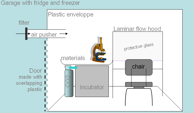
And this would be a biotech startup company, began with crowdfunding, with the writing of a detailed business plan and presentation and finding angel and venture investors, then building a top notch lab and factory, possibly making a stock market IPO, and establishing regeneration / rejuvenation / life extension clinics.
Illustration [from Wikipedia]:
The garage where two guys: Hewlett & Packard began their company
Posted Image
The somewhat related general business topics:
Bioscience/ Basement Biotech Startups
http://www.longecity...otech-startups/
Townhall/ ImmInst Goal Companies?
http://www.longecity...goal-companies/
The business idea(please do read :-) )
http://www.longecity...please-do-read/
Townhall/ Too much talk and too little action
http://www.longecity...-little-action/

Scientific literature
Some of the relevant literature:
1) Rejuvenation of aged progenitor cells by exposure to a young systemic environment. Conboy et al. 2005. Nature http://www.ncbi.nlm....pubmed/15716955
2) Loss of stem cell regenerative capacity within aged niches. ME Carlson and IM Conboy, Aging Cell(2007) 6. http://www.ncbi.nlm....pubmed/17381551
3) Molecular aging and rejuvenation of human muscle stem cells. Carlson ME et al. 2009. EMBO Molecular Medicine http://www.ncbi.nlm....pubmed/20049743
4) The ageing systemic milieu negatively regulates neurogenesis and cognitive function. Villeda et al., 2011, Nature http://www.ncbi.nlm....pubmed/21886162
5) Muscle transplantation between young and old rats: age of host determines recovery, Carlson BM and Faulkner JA, Am J Physiol. 1989 http://www.ncbi.nlm..../pubmed/2735398
6) Expansion of human cardiac stem cells in physiological oxygen improves cell production efficiency and potency for myocardial repair. TS Li et al. Cardiovasc Res (2011) 89(1). http://www.ncbi.nlm....pubmed/20675298
7) Stem cells, ageing and the quest for immortality, TA Rando, Nature, Vol 441, 29 June 2006 http://www.nature.co...ature04958.html
and other related articles, like:
Historical claims and current interpretations of replicative aging. Wright WE, Shay JW., Nat Biotechnol. 2002 Jul;20(7):682-8. http://www.ncbi.nlm....pubmed/12089552
Putative telomere-independent mechanisms of replicative aging reflect inadequate growth conditions. RD Ramirez et al., Genes Dev. 2001 Feb 15;15(4) http://www.ncbi.nlm....pubmed/11230148
Aging of Hematopoietic Stem Cells is Regulated by the Stem Cell Niche. W. Wagner et al., Exp. Geron. 2008. http://www.ncbi.nlm....pubmed/18504082
etc., i.e. further similar studies.

So, if we decide to start and do any of these above - as it is also possible to do the parts and steps in
separate subsequent projects - I imagine the whole as performed in a reward credit system (similarly to the the forum's 'Thank you points'), that is everyone who participates and contributes in any way during the course of the project, in the case of success, would be rewarded accordingly in that startup company with a proportionate share.

Edited by Avatar of Horus, 11 April 2013 - 01:24 PM.

  • like x 1

#2 kmoody

  • Guest, F@H
  • 202 posts
  • 240 â‚®
  • Location:Syracuse, NY

Posted 19 April 2013 - 04:32 PM

You have a lot of intriguing ideas here. Perhaps it would be appropriate for you to expand this into a full proposal. I would be happy to provide feedback on such a document. PM me if you would like to talk further about this.

#3 Mind

  • Life Member, Director, Moderator, Treasurer
  • 19,058 posts
  • 2,000 â‚®
  • Location:Wausau, WI
  • ✔

Posted 20 April 2013 - 12:15 PM

Also, eternaltraveler has some very similar ideas and is ready to start in the lab - just needs funding.

#4 Avatar of Horus

  • Topic Starter
  • Guest
  • 241 posts
  • 291 â‚®
  • Location:Hungary

Posted 22 April 2013 - 04:39 PM

You have a lot of intriguing ideas here. Perhaps it would be appropriate for you to expand this into a full proposal. I would be happy to provide feedback on such a document. PM me if you would like to talk further about this.

Thanx and I'd like to discuss it, so OK.

I intend to expand it, but it would be hard work for one person alone, and there are a number of currently unknown factors that are aggravating the planning procedure. Like which parts/steps would be of interest to the community to contribute and fund, and the availability of a laboratory and its inventory (like e.g. type of incubator), etc., as I'd like to focus on that portion which holds the most promise to be realized.

What I am currently doing is that I input the data of the relevant scientific literature to the computer into my BioSimulator program, to create a biological model in silico, in order to run some preliminary forecast tests.
And another one is that I'm intend to figure out the exact underlying biological processes of those regenerations/rejuvenations in the context of the biochemistry/molecular biology. Approaching it from the cell cycle, like:
from Pgs 1055 and 1102 of the Molecular Biology of the Cell, 5th Edition:
Posted Image

Posted Image

Edited by Avatar of Horus, 22 April 2013 - 04:40 PM.


#5 AgeVivo

  • Guest, Engineer
  • 2,113 posts
  • 1,555 â‚®

Posted 22 April 2013 - 04:41 PM

Avatar of Horus, I have actually started to look at how to implement some tiny portions of this at home, but with a much larger budget, and I have had the chance to accompany researchers on other portions of this. What you globally suggest seems to me like a 10 year lab program, if not what 20% of aging researchers should work on... I'm going to PM you to know you more, perhaps we can refine together and with whoever here has a good cell culture background.

Edited by AgeVivo, 22 April 2013 - 04:41 PM.


#6 Avatar of Horus

  • Topic Starter
  • Guest
  • 241 posts
  • 291 â‚®
  • Location:Hungary

Posted 22 April 2013 - 04:54 PM

Also, eternaltraveler has some very similar ideas and is ready to start in the lab - just needs funding.

Very good, the more people could be involved who sees prospect in and plans some related research the better, especially if he already has a lab, since that would largely reduce the costs. Can some details be known about his similar ideas, and the size of the required funds, etc.? I've done some searches but didn't find any details.

Since then I've also found out that some planning has been started already for a mouse research project called Age In Vivo, by a collaboration of Longecity and ILA/denigma, and it already had 6 participants. Here: http://www.denigma.d...tions/?labs=698

Possibly for a start something related to my Step 1-2 could be conducted in this cooperation and the other ones in the lab of eternaltraveler.

I'd be glad if some good scientific results could be produced, and I could help to reach that.

#7 Avatar of Horus

  • Topic Starter
  • Guest
  • 241 posts
  • 291 â‚®
  • Location:Hungary

Posted 22 April 2013 - 05:17 PM

Avatar of Horus, I have actually started to look at how to implement some tiny portions of this at home, but with a much larger budget, and I have had the chance to accompany researchers on other portions of this.

Oh, I composed my previous post (#6) before I've seen yours, so that reply partially applies to this one too.

What you globally suggest seems to me like a 10 year lab program, if not what 20% of aging researchers should work on...

Yes, my thoughts exact, actually I intended to suggest that the LE community should direct one fifth or one fourth of its life extension efforts and resources to a project like this, to see how it works, because if it proves to be as effective as the previous research results suggest, that would be a great leap forward for the cause.
I think that even it is possible that we, humanity, have already successfully conducted partial life extension, altough unknowingly and unintentionally, in the cases of the young blood into older patients transfusions.

Regarding the time frame I expect that if it would be done right, the clinical trials could be started in 3-5 years.

I'm going to PM you to know you more, perhaps we can refine together and with whoever here has a good cell culture background.

Thx and OK.

Edited by Avatar of Horus, 22 April 2013 - 05:19 PM.


#8 AgeVivo

  • Guest, Engineer
  • 2,113 posts
  • 1,555 â‚®

Posted 08 May 2013 - 04:59 PM

Leaders: for the project leader, especially for Step 1-2, I'd suggest AgeVivo

Hello, I am very honored and very pleased. It has actually been some time since I wanted to install my own cell culture lab at home, so yes, definitely I am very interested, count on me, it is exactly along my 'dream' (!) and therefore I actually have some advance on the matter:
  • For almost a year now I've been working with researchers on step 1 (not with my pseudo; those who know me well know what it is about and if you are familiar with the topic you may know with whom it is; we are making a poster this week for a conference next week)
  • in the past I had not worked much in cells but I had, so I had seen and used the required environment for a cell culture lab
  • I also within the lab I had created some clean room sections in a very cheap way so I have some experience that is good for a DIY lab.
The overal steps in the first post if this thread are too big I think for a foreseable LongeCity grant, so as discussed with Avatar of Horus we would create a project a DIY cell culture biohack lab toolkit with one application in life extension . It means that
  • 1. with the help of others in LongeCity and elsewhere we would try to build a cell culture DIY lab in my garage, in a cheap, easy and reproducible way, as much as possible (balance to be found between the 3 caracteristics)
  • 1'. We would document it and try to make it as easy as possible for willing persons (a priori, non researchers and retired researchers) to invest a few thousand $ and do the same in their garage
  • 2. We would do a first life extension application around the ideas expressed above (of course you know our longevity dedication so you know we'll go much further if we can; especially cell culture+mouse applications)
  • 2'. We would document it and try to make it as easy as possible for willing persons (esp researchers) to reproduce it
  • A LongeCity grant would pay for the materials. Before spending many hours to go in fine details, Does this seem foreseable for LongeCIty directors?
We have discussed it with Avatar of Horus these last days and here is some first visions:

Here is the proposed installation:
DIYlab_v1.png
That image represents what I think is the minimal version of a cell culture DIY bio lab: the clean area (in the plastic enveloppe) would be 2 by 1 meters on the floor. There should also be a fridge+freezer next to it. It would be for one person inside at a time. I can make a bigger installation but this has the merit I think to fit for as many persons/houses as possible.

Note1: The plastic enveloppe + filtered air pusher allow to work in a clean environment. In a normal lab to avoid contamination of cells by bacterial, a cell culture room is indeed kept very clean AND laminar flow hoods are used.
Note2: What we propose can absolutely not be used to manipulate dangerous things (I would not put such a thing online, to avoid undesirable persons do undesirable things)

Our very first estimation of the costs ranges between 5k$ and 10k$ :
- laminar flow hood about 2.5 k€
- 37°C CO2 incubator about 2 k€ (CO2 bottle does not cost much but the pressure regulator does)
- centrifuge + inversed microscope about 1 k€
- small materials 1) for the environment (such as plastic,air pusher with filters, metal bars to create enveloppe..), 2) tools such as a pipetteman 3) sterile consumable (such as 1/5/10 ml pipettes, petri plates and eppendorfs), chemicals (such as trypsine, foetal bovine serum,...) : 1 or 2 k€

Note1. a big thank to friend researchers and to people on http://webchat.freen...=##hplusroadmap who advised on this.

The life extension application now needs to be defined, Avatar of Horus is working on it.
There well will of course be restrictions. Eg a FACS to sort cells costs more than 20 k$...

*******************************************************************************************************************
General note: anyone here with cell culture experience: suggestions are welcome.
*******************************************************************************************************************

Edited by AgeVivo, 08 May 2013 - 05:07 PM.

  • like x 1

#9 Avatar of Horus

  • Topic Starter
  • Guest
  • 241 posts
  • 291 â‚®
  • Location:Hungary

Posted 08 May 2013 - 07:20 PM

You have a lot of intriguing ideas here. Perhaps it would be appropriate for you to expand this into a full proposal...

I intend to expand it...


I have completed the first round of the expansions, and here I present these in a couple of subsequent posts in the following days as I've finalized the texts.

The expansions are to: the research, science, medicine, and the DIYBio tech/equipment, and the business parts.
The intention of these is to give a broader scientific background and more accessory informations, and to serve as the base for the another round of expansion: the working out of the concrete and detailed research experiment protocols, which will be the foundations for the therapies.

A preliminary description of the planned whole medical/biotech procedure, so it would be eventually something like this:

10-100 ml blood is taken,

separating the blood cells and the plasma/serum,

thus the various cells' number would be several millions or so, including a number of adult/somatic stem cells, like hematopoietic stem cells/HPSC. Possibly combining this with some bone marrow and adipose/fat tissue harvesting too, for retrieve more HPSCs and other stem cells, like mesenchymal stem cell/marrow stromal cells/MSCs, but this possibly won't be needed. Then separating the cells as well to the different types.

With these installing the cell and tissue culture setup for culturing the cells on the serum medium, then first creating those cells/tissues which are needed for the autologous bio-artificial blood producing, like the blood cells from the HP/blood stem cells and the serum,

then with these: applying the young blood and serum therapy, and also with this bio-artificial serum supply culturing the other tissues for the 'young backups'.

As I've mentioned already all this eventually would be done by an automatic machine system, to which the blood would be inserted, along with the necessary food based nutrients.


And now about the expansions - all of them are, to a degree, interrelated to each other -,
the structure and subjects of these will be:
- first, some more words to the bussiness part, and
- a general overview of the whole aging process, in a simplified way,
- then some examples for this in the form of age-related degenerative conditions,
- then some of the means of redeeming and compensating them: informations about a number of already started human serum based regenerative therapies and treatments,
- then some description and expansion to the low oxygen and
- human serum based cell culturing techniques, and for the checking procedure,
- and finally a general scientific description on some biological processes that are partially underlying all these aging and regeneration / rejuvenation effects on the molecular biology level.

And about the aim:

... actually I intended to suggest that the LE community should direct one fifth or one fourth of its life extension efforts and resources to a project like this, to see how it works, because if it proves to be as effective as the previous research results suggest, that would be a great leap forward for the cause.
Regarding the time frame I expect that if it would be done right, the clinical trials could be started in 3-5 years.

an example for this, one of the "previous research results":
a quote from the abstract of [Sci3]:
"... restored ‘youthful’ myogenic responses to satellite cells from 70-year-old humans, rendering them similar to cells from 20-year-old humans. These findings strongly suggest that aging of human muscle maintenance and repair can be reversed..."
"70 year-old to 20 year-old" what's this if not impressive? :) I will get back to this later in the science section.

Note: the posts may be long, but all is relevant, and also wherever relevant there will be quotes from the sources, because I want that as much info as possible be together and in one place.

So stay tuned!

Edited by Avatar of Horus, 08 May 2013 - 07:23 PM.


#10 kmoody

  • Guest, F@H
  • 202 posts
  • 240 â‚®
  • Location:Syracuse, NY

Posted 08 May 2013 - 07:32 PM

You may consider structuring your proposal as a Google Doc or similar and sharing the link rather than making this thread too dense.

I would also suggest you focus on just one of your "steps" and define the research plan for that step in excruciating detail (background / rationale for why you want to do it, specific outcomes you will assess, a methods section that describes how the techniques will be performed, a facility section that identifies what equipment / infrastructure you need, comprehensive budget for salaries, equipment, reagents, and consumables [I would leave out dollar figures for now because much of what you need may be available through the community]).

I think that any single one of your steps would be remarkably ambitious, certainly in terms of the level of funding that Longecity could provide. That does not mean they are bad ideas in principle. Rather, you should consider picking just one, make it work, and use that data to justify your request for additional funding from Longecity and/or other sources.
  • like x 1

#11 kmoody

  • Guest, F@H
  • 202 posts
  • 240 â‚®
  • Location:Syracuse, NY

Posted 08 May 2013 - 07:38 PM

SENS Foundation has a template available for what such a document may look like. See http://www.sens.org/...t_Proposal.pdf.
  • like x 1

#12 Avatar of Horus

  • Topic Starter
  • Guest
  • 241 posts
  • 291 â‚®
  • Location:Hungary

Posted 09 May 2013 - 06:03 AM

You may consider structuring your proposal as a Google Doc or similar and sharing the link rather than making this thread too dense.

I would also suggest you focus on just one of your "steps" and define the research plan for that step in excruciating detail (background / rationale for why you want to do it, specific outcomes you will assess, a methods section that describes how the techniques will be performed, a facility section that identifies what equipment / infrastructure you need, comprehensive budget for salaries, equipment, reagents, and consumables [I would leave out dollar figures for now because much of what you need may be available through the community]).

I think that any single one of your steps would be remarkably ambitious, certainly in terms of the level of funding that Longecity could provide. That does not mean they are bad ideas in principle.

Thanx for the suggestions.

These texts will be in a summary style, and not so extremely long, in total about twice the size of my first post, and they will be separated into 6 or so posts.

First some general info:
in my thinking this project idea - as I wrote in the initial post - is for life extension DIYBio experiments of the community science type, and so it's actually just "an initial outline" and a bunch of suggestions "for consideration, discussion and expansion, modification, etc., as I imagine the whole thing as a community project, that is the members of the community do the further planning and refinements, etc, then a couple of people do this or that part of it."

And the purpose of this topic and these texts is to present: how it can be done, what equipment is needed for it, which literature it is based on, what has already been carried out and accomplished, why it may be scientifically relevant and useful, and so on; and as I've written here in the 9th post: to be a "... base for the another round of expansion: the working out of the concrete and detailed research experiment protocols...".

And this latter would and will be that detailed proposal document you are suggesting.

So, regarding this:
with AgeVivo we've already started to work out a minimalist setup for a concrete proposal. I expect this document would be ready in a couple of weeks.
The aim of it is twofold, it would be:
a 'DIYBio lab toolkit', a guide for starting a lab for life extension experimenting, with listing the equipments needed, the experimental protocols, etc., to be available to the interested other peoples of the LE and DIYBio communities around the world, to start this type of research;
and for the real establishment of such a lab, for a concrete life extension experiment based on cell culturing.

As I've already also written: the main costs are for the equipments, like the laminar flow hood, cell incubator, etc., the ones that are in my posts and also AgeVivo started to list above. This too will be completed.
And once these have been set up, the experiments can be started, and can be continued and extended later and on the fly, with already lower costs, according to one's liking and the availability of further funding and interest and will and such.

And this latter is exactly the reason too why I've designed and written it in the starting post that way, that it can be performed step-by-step.

But actually the eventual aim of this approach is that the automatic biotech system, which would be engineered based on the the results of those smaller conducted experiments' elements, would be - in my current conception - a self-sustaining system, that after its set up, only needs some electricity and a nutrient supply, and with these the needed tissues would grow themselves out on their own from an initial cell culture, like a whole organism or a human body from one cell, and these would continuously produce the young blood and 'youth serum' and the young cells and tissues for the periodical rejuvenation of the adult body.

Rather, you should consider picking just one, make it work, and use that data to justify your request for additional funding from Longecity and/or other sources.

Actually in my plan just that would be done, but with the angel and venture capitalist, after some initial results that can be presented as a proof of concept have been achieved. But right, possibly it would be good to be applied here too.

SENS Foundation has a template available for what such a document may look like. See http://www.sens.org/...t_Proposal.pdf.

Thanx for this template.
A minor note: to me the link gives a not found error, because of the ending period; so the good link is this: http://www.sens.org/...nt_Proposal.pdf

#13 Avatar of Horus

  • Topic Starter
  • Guest
  • 241 posts
  • 291 â‚®
  • Location:Hungary

Posted 10 May 2013 - 07:03 AM

first, some more words to the bussiness part


Another related possibility to that automatic machine system would be the manufacturing of a 'personalized health kit', what would be similar to a first aid kit and cooler box, which would contain preproduced autologous biomaterials, like e.g. a liter blood, and a couple millions of various healthy cells for the fast tissue repair in the case of injuries.
Posted Image

and
to this: "finding angel and venture investors" two older blog articles, which are nicely presenting some of the points.
(I quote them in case they'd disappear from the net, like two of their links already):

"Status of Research on Human Aging
Tuesday, July 01, 2008

Longevity is the new alternative energy
With $10m quickly raised by the Methuselah Foundation, VCs just beginning to see the opportunity and continually soaring healthcare costs, the longevity market could easily become as big as the alternative energy/climate change solutions market has become now.

Longevity research status
Grossly generalizing, the main focus in aging research is figuring out how to get processes that already occur, in the young and in cancer for example, to occur at other times, in the old. The optimum approach may include both reverse engineering and forward engineering in the form of synthetic biology as has been successful in other biological research areas like gene synthesis.

Aging is multidisciplinary, comprising at minimum the study of stem cells, immunology, cancer, DNA damage, tissue engineering, genetic engineering, regenerative medicine and micronutrients.

A comprehensive collection of anti-aging research findings was presented at the Aging 2008 conference June 27-29 at UCLA. The current developmental stage of aging research is early, perhaps in the second inning. Groundwork is being laid, phenomena are being documented, understanding of general mechanisms is sought, existing processes are being enumerated and early cycles of testing have begun primarily on flies and mice.

The seven primary causes of aging are DNA mutations in the cell nucleus and mitochondria, junk that builds up inside and outside cells, cells sticking together and cell loss and death. These are described at length, together with potential solutions, in aging research pioneer Aubrey de Grey’s book, Ending Aging and in the journal Rejuvenation Research. De Grey’s organization, the Methuselah Foundation, provides grants to anti-aging researchers. Some of the freshest thinking so far has included biomedical remediation, therapeutic organisms purpose-catalyzed in the body and the possibility of removing the overly-prone-to-damage mitochondrial DNA.

Generalized summary of Aging 2008 research findings:

- Applying (non-individual specific) substances from the young to the old appears to work
- With aging, not only does "good stuff" (cells, processes, etc.) decline but "bad stuff" also arises
- The quality of the biological environment facilitates or inhibits activity and repair
- Treatments may be most effective when begun in youth or middle age
- The goal is to extend healthspan not just lifespan

DIY biohacking and the cocktail problem
Every bit as interesting as the scientific talks were the informal discussions of the wide range of interventions, treatments, supplements and other anti-aging remedies in use by conference participants.
The cocktail problem is how multiple remedies taken in concert may be impacting each other. Never has there been a market with such demand and so few offerings as for anti-aging remedies.
" Source: http://futurememes.b...uman-aging.html

I will get back later also to this ImmInst/Longecity co-sponsored Aging 2008, aka UABBA, conference in
the science section.

"Next big VC market: life extension?
Sunday, July 20, 2008

Life extension is a growing market and could be the next significant industry targeted by Venture Capitalists and private investment as alternative energy and clean tech eventually wane. The opportunity is made obvious by continuous soaring costs in the world’s largest industry, healthcare, unfunded Medicare type liabilities in every industrialized country, and the demographic aging of populations and below replacement fertility rates together with massive demand and willingness to spend on longevity remedies.

What is the Life Extension Market?
The life extension market is the commercialization of scientific findings from stem cell, immunology, cancer, regenerative medicine and other areas of research. The research linkage between products and research will hopefully become stronger and more standardized. For example, many longevity remedies available today claim scientific support. This research could and should be linked to the products online (23andme is a nice example of research linkage) so consumers and other interested parties can research the products themselves. Bloggers and other independent intermediary watchdogs could synthesize the scientific research and confirm or deny the product claims.

Longevity Docs Needed
It is not clear that traditional physicians will be those prescribing longevity remedies. Specialist longevity docs are needed and will likely arise and market themselves as such, there are a few examples of this today. Most traditional physicians do not currently have expertise in new areas such as longevity and personalized genomics, or the enhancement and prevention vs. cure mindset.

Supplements, Hormones and Enzymes
The first step in life extension treatments is supplements, ranging from a daily multivitamin to the 200 or more supplements per day taken by futurist Ray Kurzweil. The next step is hormone and enzyme
replacement therapies, which must generally be overseen by a physician. A variety of treatments have
been undertaken per the shifting legal climate, not everyone wanting to be restored to the hormonal levels of their twenties and other reasons.

Longevity Social Network
It would be great to have a health social network (HSN), like PatientsLikeMe and CureTogether for the longevity community. First, people could share the different interventions they are trying. Second, they could upload their ongoing bio-marker test data into an aggregated electronic health record, similar to what Google Health is contemplating, to track and possibly share the impact of the interventions. Third, companies with research and therapies targeting this market could contact an aggregated group to propose field studies, clinical trials and offerings. For example, the 23andme Parkinson’s community has been contacted for such research.
" Source: http://futurememes.b...-extension.html

Also there will be another business related idea presented later in the serum based therapy post.

#14 AgeVivo

  • Guest, Engineer
  • 2,113 posts
  • 1,555 â‚®

Posted 12 May 2013 - 01:16 AM

Dear Avatar of Horus,
When I read your posts above I see some true goldmines (I am very impressed by the links between specific parts of your readings); to me, they are located at dense, unexpected places like the last image of post #4, so I just prefer to say it in advance for your 6 posts: Perhaps you want to put in red where you want to go, in blue the key technical results you are based on... And in any case it might be good to then make google docs out of them that volunteers may want to sligthly restructure (they will learn interesting knowlege and connections at the same time!). Waiting to read you!

Edited by AgeVivo, 12 May 2013 - 01:17 AM.

  • like x 1

#15 Avatar of Horus

  • Topic Starter
  • Guest
  • 241 posts
  • 291 â‚®
  • Location:Hungary

Posted 12 May 2013 - 09:58 PM

Dear Avatar of Horus,
When I read your posts above I see some true goldmines (I am very impressed by the links between specific parts of your readings); to me, they are located at dense, unexpected places like the last image of post #4, so I just prefer to say it in advance for your 6 posts: Perhaps you want to put in red where you want to go, in blue the key technical results you are based on... And in any case it might be good to then make google docs out of them that volunteers may want to sligthly restructure (they will learn interesting knowlege and connections at the same time!). Waiting to read you!

Thanx for the appreciation and for the suggestions, I intend to use those color things later and also create a google doc, but these 6 posts just provide some background infos and examples to help the general overview of the whole thing. Here is the second post, for the:

- a general overview of the whole aging process, in a simplified way,

So here is a general overview of aging, from the viewpoint of the growth:

All this extracellular environment thing can make sense as well in the context of the developmental biology. That is if the aging is simply seen as a process of change in the size and state of the body, and its organs and tissues, i.e. the health and number of the cells:

in this, 3 phases can be discerned, the:
young, adult, old
and these corresponding to
growth, balance, degradation.

young: growth/development, increasing in cell numbers and functions, overally healthy state
adult: balance in cell numbers and functions, health maintained
old: decrease in cell numbers and functions, health diminishes

An illustration from the Internet:
Posted Image
Source: http://saplinz.wikis...uman_Growth.jpg

The exact mechanisms that underlying the whole aging process are not known currently, whether is it a 'premediated' genetic program or just accumulating damage, that retards the normal functioning, or a mix of these. But the growth course is a pre-programmed one and this program determines the body's growth, its size, etc. And the program is encoded in the genome. And so the whole develepment of the body is controlled by a continuous interplay, starting from the womb and zygote, between the cells' internal genetics and external environment. But these also are interconnected because the extracellular environment is also depends on the genetic program, which determines the molecular products and secretions of the cells, which are interpreted by the other cells, and so on. Like the general levels of the growth and proliferation and differentiation molecules in the blood and other extracellular fluids.

The point here is that in what degree the normal functioning of the body and its cells processes are behind those accumulated damages.
And so the question arises: is this healthy environment enough for the constant health maintenance or not in itself, but there is a need also for the growth related calibrations of this environment. That is what can be considered as the 'healthy extracellular environment', and what are the normal turnover of the cells and their numbers in the above second phase.

As I've written here: quote from the #1 post:
"Step 4
The point of this step is to try to maintain an as much as possible optimal, constant young and healthy in vivo environment, which was determined with the help of Step 3, for all of the cells and tissues of the body, through the manipulation of the blood, that is an indirect intervention into the interstitial fluid in the extracellular space. I.e. this route would be the therapeutic pathway. Or with a direct one later with
microneedles and such."

There are two distinct possible conditions here: one is the stopping the aging process, that is halting the degrading changes in the healthy adult state, and then continouosly maintaining this, by removing e.g. the toxins and the misfolded proteins, and such;
and the second is the regeneration, that is to restore the tissues' cell losses if they had already happened, in the case of aged peoples.
So the point and the aim may be that the development regulating molecules' levels need to be calibrated to the levels of the 20-40 years old age.
That is the Reprogramming of the aging process or Biohacking the aging process.

This change process can be illustrated well with the diagram of human growth hormone changes; note, however, that this is just a sub-part of the whole extra cellular control thing:
Posted Image
source: Human growth hormones for anti aging, Published February 4, 2013
http://www.thenutrit...anti-aging.html
and
Posted Image
source: How to increase HGH (Human Growth Hormone), February 12, 2013
http://mineposting.b...th-hormone.html

So in other words, it is possible that in this therapy we may also need to maintain that level of the extracellular environment that controls the cells and tissues as if they were in the 20-30 or 30-40 years old.
First with the young serum and its elements, and later if possible identify the exact molecular factors, as this was mentioned in some quotations of that Guardian article, given the end of the #1 post.

For example the embryonic stem cells may have the ability to secrete molecules that can control and program the environment to support growth. As this was illustrated in the first post, in the figure from [Sci2/ Fig.6].

To these above are related some presentations of the mentioned UABBA 2008 conference:
A quote from:
t2) Bioscience/ Young Blood Reverses Signs of Aging in Old Mice http://www.longecity...ng-in-old-mice/
[t2/4]:

This type of effect was also discussed at UABBA, a conference Imminst sponsored 2 years ago. I.M. Conboy review on stem cell signalling pathways, Leanne Jones, age-related changes to the stem cell niche.

So this news was not really anything new to me and the Harvard research did not find the main driving force behind the rejuvenation of the old mice, just more speculation. I suspect it involves several signaling factors working in concert.

and the quotes from the given links, from the topic 'UABBA Conference Coverage':

I.M. Conboy "Aging of signal transduction in stem cells"

She described a process for up-regulating stem cell activity in aged muscle tissue. "We don't lose the capacity to regenerate muscle because of the lack of muscle stem cells, but because of the deregulation/degradation of signaling pathways.

Apparently transplanting young stem cells into old tissue does not lead to rejuvenation. Instead the young stem cells take on characteristics of the aged tissue.

"Aging can be described as a negative force exerted by old differentiated niches on stem cells".

She also mentioned something about ESCs having the ability to resist the "negative force" exerted by old tissue, whereas adult stem do not. (I can't be sure on this point because she has a thick accent and I couldn't follow closely).

She says TGF-beta is one of the main age related inhibitory culprits - one of the main negative forces. TGF-beta

Leanne Jones: "Age-related changes changes to stem cells and the stem cell niche"

She described the process by which the stem cell niche influences stem cell production and thus tissue repair and homeostasis.

Reduced signaling within the stem cell niche contributes to decreased stem cell function. Forced (unnatural) expression of some signals (specifically unpaired - upd) stops the decrease of stem cell function and the loss of stem cells and niche cells.

She suggests that regenerative medicine based on transplanting stem cells into old tissue will have to consider the signals from the niche as well. In order for young stem cells to work in old tissue you may need to also transplant the stem cell niche.

Is the stem cell niche signalling genetically regulated? A question her team is still investigating.

Do changes in the niche select for self-renewing stem cells that can operate independent of the niche - ie. cancer stem cells? Another open question.
...


#16 AgeVivo

  • Guest, Engineer
  • 2,113 posts
  • 1,555 â‚®

Posted 13 May 2013 - 03:50 AM

"Biohacking the aging niche process" in that we stimulate stem cell activity -- isn't growth hormone supplementation already a good solution to that approach? (I haven't checked that growth hormone does it; just a question and a guess; I imagine the answer is in the literature). If so, -- and yet, growth hormone isn't in fact the solution to aging.

If so, shouldn't "biohacking aging" also be about making room for regeneration? I am in particular thinking about the lymphocyte pool, that saturates the immune system activity when it becomes too big. Also about resistant senescent cells that accumulate and produce inflammation and other long term poisoning factors?

<<very theoretical thought, that might be wrong I don't know: that could explain caloric restriction: such accumulations/saturations rapidly become a burden for the body when the body stops growing, such that under CR the body having produced less has less of such a general burden and has more liberty to use its normal functions like lymphocyte expansion>>

#17 Avatar of Horus

  • Topic Starter
  • Guest
  • 241 posts
  • 291 â‚®
  • Location:Hungary

Posted 21 May 2013 - 08:51 AM

"Biohacking the aging niche process" in that we stimulate stem cell activity -- isn't growth hormone supplementation already a good solution to that approach? (I haven't checked that growth hormone does it; just a question and a guess; I imagine the answer is in the literature). If so, -- and yet, growth hormone isn't in fact the solution to aging.

If so, shouldn't "biohacking aging" also be about making room for regeneration? I am in particular thinking about the lymphocyte pool, that saturates the immune system activity when it becomes too big. Also about resistant senescent cells that accumulate and produce inflammation and other long term poisoning factors?

<<very theoretical thought, that might be wrong I don't know: that could explain caloric restriction: such accumulations/saturations rapidly become a burden for the body when the body stops growing, such that under CR the body having produced less has less of such a general burden and has more liberty to use its normal functions like lymphocyte expansion>>

Note: I didn't planned this text and its topic in the course of the mentioned (in post #9) 6-7 initial overview posts, only at a later stage, but here I've felt the need for some more clarification and explanation. So here it is:

In the aspect of this approach -
it's important to note that all this is theoretical, that is science suggested theories and hypotheses, which need to be tested experimentally -
aging is the aftermath of the develepmental phase and program, and the goal of the latter is the reproduction of the individuals and the conservation of the species, and therefore the body / genetic program codes for the maintenance only until about the twice of the reproductive age, that is for example around year 40 in humans, because of the offsprings' need for parental care. And this was evolutionary developed/selected to be such, or - to be said this too - was, along with the evolution, designed and created so by a powerful intelligence.

And so in the context of this, aging can be seen as the unregulated afterphase of the body growth/development phase, and thus its consequence, that of the lack of the further control of maintenance, is the degradation.

Describing physiologically aging is the differences or the structural and functional changes from the young to aged tissues/organs. And the original healthy state, that is the homeostasis needs to be maintained with the damages repaired and losses restored.

So, the above 3 phases in other words:
generation, maintenance, and degeneration,

the generation is the -genesis phase: like e.g. embryogenesis, organogenesis, morphogenesis;
the maintenance is the homeostasis; and
the degeneration is e.g. the degenerative diseases, and from this follows that the solution to this is the regeneration, that is, as the word itself too tells it, "generate again".
And for the (re)generation new cells are needed, the 'genesis', and those are created by mitosis, more precisely with cell division, and this is initiated by the mitogens, as was mentioned in the #4 post.

As I've written here:

"The point here is that in what degree the normal functioning of the body and its cells processes are behind those accumulated damages.
And so the question arises: is this healthy environment enough for the constant health maintenance or not in itself, but there is a need also for the growth related calibrations of this environment. That is what can be considered as the 'healthy extracellular environment', and what are the normal turnover of the cells and their numbers in the above second phase."


An illustration for this, from the post #1's [Sci7]:
Posted Image
and some other literature:

Apoptosis in the pathogenesis and treatment of disease
Thompson CB.
Howard Hughes Medical Institute, Department of Medicine, Chicago, IL
Science. 1995 Mar 10;267(5203):1456-62.
http://www.ncbi.nlm..../pubmed/7878464
Abstract
In multicellular organisms, homeostasis is maintained through a balance between cell proliferation and cell death. ...

Polarity in stem cell division: asymmetric stem cell division in tissue homeostasis
Yamashita YM, Yuan H, Cheng J, Hunt AJ.
Cold Spring Harb Perspect Biol. 2010 Jan;2(1):a001313. doi: 10.1101/cshperspect.a001313.
Life Sciences Institute, Center for Stem Cell Biology, University of Michigan, Ann Arbor, USA.
http://www.ncbi.nlm....pubmed/20182603
Abstract
Many adult stem cells divide asymmetrically to balance self-renewal and differentiation, thereby maintaining tissue homeostasis. Asymmetric stem cell divisions depend on asymmetric cell architecture (i.e., cell polarity) within the cell and/or the cellular environment. In particular, as residents of the tissues they sustain, stem cells are inevitably placed in the context of the tissue architecture. Indeed, many stem cells are polarized within their microenvironment, or the stem cell niche, and their asymmetric division relies on their relationship with the microenvironment. Here, we review asymmetric stem cell divisions in the context of the stem cell niche with a focus on Drosophila germ line stem cells, where the nature of niche-dependent asymmetric stem cell division is well characterized.

And so this tissue homeostasis is maintained by the stem cells:
for example: the text of the picture of Figure 1 of post #1, from [Sci7]:
Posted Image
and it is controlled by stem cell niche locally.
And more generally by - that is the regulation itself, one of its main form, is composed of - the regulationary molecules in the blody fluids. Like e.g. the growth factors and the endocrine system's hormones. And the levels of these control molecules are changing during the aging process.
And a part of the 'Reprogamming and Biohacking the aging process' is (from the above quote of mine) the "growth related calibrations of this environment". That is the control of the levels of these and other molecules: "So the point and the aim may be that the development regulating molecules' levels need to be calibrated to the levels of the 20-40 years old age.". Whose effects will be tested in the cell culture step, and on already aged tissues and on animals, and later also the production will be with that autologous biotech ex-vivo in-vitro cell / tissue culture system. And so with this system we can hit two birds with one stone: that is creating the necessary cells and tissues, by which we can determine and microcalibrate the molecular regenerative coctail's composition and also test it ex vivo on generated tissues, and produce the constituent biomolecules themselves.

Another part of the 'Reprogamming/Biohacking the aging process' is the "healthy environment ... for the constant health maintenance", which is related to the protein homeostasis, about this I will write some words in my next post.

quote from: http://en.wikipedia....ndocrine_system
"The endocrine system is the system of glands, each of which secretes different types of hormones
directly into the bloodstream..."
For example: the hypothalamus produces growth-hormone-releasing hormone, GHRH, after which the pituitary gland releases growth hormone.

A similar recent related finding is here: the topic of Bioscience/Aging Theories: Hypothalamus Key to
Aging? : http://www.longecity...s-key-to-aging/

And I used the growth hormone and its picture as an example and an illustration for the whole developmental molecular regulation thing, of which, as I wrote, the GH is only a part: "This change process can be illustrated well with the diagram of human growth hormone changes; note, however, that this is just a sub-part of the whole extra cellular control thing:"

Some other literature related to this:

Hormonal regulation of longevity in mammals
Brown-Borg HM.
Ageing Res Rev. 2007 May;6(1):28-45.
Department of Pharmacology, Physiology and Therapeutics, University of North Dakota School of
Medicine and Health Sciences
http://www.ncbi.nlm....pubmed/17360245
Abstract
Multiple biological and environmental factors impact the life span of an organism. The endocrine system is a highly integrated physiological system in mammals that regulates metabolism, growth, reproduction, and response to stress, among other functions. As such, this pervasive entity has a major influence on aging and longevity. The growth hormone, insulin-like growth factor-1 and insulin pathways have been at the forefront of hormonal control of aging research in the last few years. Other hormones, including those from the thyroid and reproductive system have also been studied in terms of life span regulation. The relevance of these hormones to human longevity remains to be established, however the evidence from other species including yeast, nematodes, and flies suggest that evolutionarily well-conserved mechanisms are at play and the endocrine system is a key determinant."


Currently the topic of growth hormone and aging/longevity is controversial and inconclusive.
For example:

Growth hormone and aging
Bartke A.
Endocrine. 1998 Apr;8(2):103-8.
Department of Physiology, Southern Illinois University School of Medicine, Carbondale 62901, USA.
http://www.ncbi.nlm..../pubmed/9704566
Abstract
Although age-related decline in plasma growth hormone (GH) levels is well documented, the possible role of GH in the control of aging is controversial. Overexpression of GH in transgenic mice is associated with reduced life expectancy and numerous symptoms of premature aging. Ames dwarf mice with hereditary GH, prolactin, and thyrotropin deficiency live much longer than their normal siblings. In contrast to these indications that GH may accelerate aging, some physiological changes in the elderly resemble symptoms of GH deficiency and can be corrected by GH replacement. It is suggested that these seemingly contradictory observations are related to the dose-response characteristics of GH action, and to negative correlation between body size and life expectancy within a species. Physiological mechanisms linking plasma GH levels and body size with aging remain to be identified.


At this point it can be mentioned that the one of the Methuselah Mouse Prize winning was achieved by the manipulation of the growth hormone thing, with a "Growth Hormone Receptor Gene Knockout transgenic mouse"; the second one on the MPrize web page:
http://www.mprize.or...j_mprize_record

Life extension in the dwarf mouse
Bartke A, Brown-Borg H.
Curr Top Dev Biol. 2004;63:189-225.
Geriatrics Research, Department of Medicine, Southern Illinois University School of Medicine, Springfield,
Illinois, USA.
http://www.ncbi.nlm....pubmed/15536017
Abstract
Ames dwarf mice and Snell dwarf mice lack growth hormone (GH), prolactin (PRL), and thyroid-stimulating hormone (TSH), live much longer than their normal siblings, and exhibit many symptoms of delayed aging. "Laron dwarf mice," produced by targeted disruption of the GH receptor/GH-binding protein gene (GHR-KO mice), are GH resistant and also live much longer than normal animals from the same line. Isolated GH deficiency in "little" mice is similarly associated with increased life span, provided that obesity is prevented by reducing fat content in the diet. Long-lived dwarf mice share many phenotypic characteristics with genetically normal (wild-type) animals subjected to prolonged caloric restriction (CR) but are not CR mimetics. We propose that mechanisms linking GH deficiency and GH resistance with delayed aging include reduced hepatic synthesis of insulin-like growth factor 1 (IGF-1), reduced secretion of insulin, increased hepatic sensitivity to insulin actions, reduced plasma glucose, reduced generation of reactive oxygen species, improved antioxidant defenses, increased resistance to oxidative stress, and reduced oxidative damage. The possible role of hypothyroidism, reduced body temperature, reduced adult body size, delayed puberty, and reduced fecundity in producing the long-lived phenotype of dwarf mice remains to be evaluated. An important role of IGF-1 and insulin in the control of mammalian longevity is consistent with the well-documented actions of homologous signaling pathways in invertebrates.

and

The endocrine regulation of aging by insulin-like signals
Tatar M, Bartke A, Antebi A.
Science, 2003. Feb 28;299(5611):1346-51.
Department of Ecology and Evolutionary Biology, Brown University, Providence, RI 02912, USA.
http://www.ncbi.nlm....pubmed/12610294
Abstract
Reduced signaling of insulin-like peptides increases the life-span of nematodes, flies, and rodents. In the nematode and the fly, secondary hormones downstream of insulin-like signaling appear to regulate aging.
In mammals, the order in which the hormones act is unresolved because insulin, insulin-like growth factor-1, growth hormone, and thyroid hormones are interdependent. In all species examined to date, endocrine manipulations can slow aging without concurrent costs in reproduction, but with inevitable increases in stress resistance. Despite the similarities among mammals and invertebrates in insulin-like
peptides and their signal cascade, more research is needed to determine whether these signals control aging in the same way in all the species by the same mechanism.


However, regarding this reduced GH it is also important to mention that what I was talking about above and this latter are the different ends of the (partially) same thing, because, in my current view, this life extension was achieved by slowing down or reducing the growth phase, that is for instance the speed and quantity of the metabolism, without the repair and regeneration thing, and so the damage was accumulating at a slower pace, and thus the same pathological level was reached in a longer time, so this indeed was increased longevity, but, following from its nature, just definite life extension.

The one that is more similar to the topic of my above writings is this:
it is also mentioned in the Wikipedia's Life Extension page:
http://en.wikipedia....mone_treatments
"An early study suggested that supplementation of mice with growth hormone increased average life expectancy."

Effects of long-term, low-dose growth hormone therapy on immune function and life expectancy of mice
David N. Khansari, Thomas Gustad
Mechanisms of Ageing and Development, Volume 57, Issue 1, January 1991, Pages 87–100
Department of Veterinary Science/Microbiology, North Dakota State University, Fargo, ND 58105 U.S.A.
http://www.ncbi.nlm..../pubmed/2002700
http://www.sciencedi...04763749190026V
Abstract
We have studied effects of long-term, low-dose growth hormone therapy on the immune function and life expectancy of Balb/c mice. Sixty male Balb/c mice were aged up to the time when they started showing signs of senescence and casual death (deaths started when they became 17 months old). The aged mice were divided into two groups of 26 mice each. One group received growth hormone (309 ug/mouse) subcutaneously twice a week for 13 weeks. The control group received an equal volume of saline for the same period. During this treatment period, 16 control mice died (61%) whereas only 2 of the hormone-treated micef died (7%). Four mice from each group were killed and immunological functions of splenocytes were evaluated. Hormone-treated mice had higher stimulation indices for pokeweek mitogen but not for Concanavalin-A. Total IgG production was decreased but IL-2, IL-2 and TNF production was increased. After a lag period of 4 weeks, growth hormone therapy was continued for another 6 weeks. One of the growth hormone treated mice died while the control group no longer existed. Splenocyte functions of the growth hormone treated mice were compared to those of young mice. The results showed no significant difference between cytokine production (IL-1, IL-2, TNF and IgG) in the young and the hormone treated groups. Stimulation induced by concanavalin-A and pokeweed mitogen however, was higher in the young group than the old group. The mortality curve obtained suggests that long-term low-dose growth hormone treatment prolongs life expectancy.


To the "making room for regeneration" part I will answer in my next post.

Edited by Avatar of Horus, 21 May 2013 - 09:02 AM.


#18 Avatar of Horus

  • Topic Starter
  • Guest
  • 241 posts
  • 291 â‚®
  • Location:Hungary

Posted 21 May 2013 - 08:57 AM

...
If so, shouldn't "biohacking aging" also be about making room for regeneration? I am in particular thinking about the lymphocyte pool, that saturates the immune system activity when it becomes too big. Also about resistant senescent cells that accumulate and produce inflammation and other long term poisoning factors?

<<very theoretical thought, that might be wrong I don't know: that could explain caloric restriction: such accumulations/saturations rapidly become a burden for the body when the body stops growing, such that under CR the body having produced less has less of such a general burden and has more liberty to use its normal functions like lymphocyte expansion>>

To the "making room for regeneration" part I will answer in my next post.

This depends on the state of degeneration of the tissues. Because in this state the cells' numbers are already can be smaller. So one initial regenerative intervention can be made. And later the control of this in my thinking will be with the autologous young blood's cells: quote from my #1 post:

4) ...
This step may also include an immune system repairment...

I will discuss the degenerative diseases in the next course post and some more the immune system in the young environment/serum based therapies post.

And to the "protein homeostasis" an example:

Delaying aging and the aging-associated decline in protein homeostasis by inhibition of tryptophan degradation
Annemieke T. van der Goot et al.
PNAS vol. 109 no. 37 September 11, 2012
http://www.pnas.org/.../14912.abstract
Abstract
Toxicity of aggregation-prone proteins is thought to play an important role in aging and age-related neurological diseases like Parkinson and Alzheimer’s diseases. Here, we identify tryptophan 2,3-dioxygenase (tdo-2), the first enzyme in the kynurenine pathway of tryptophan degradation, as a metabolic regulator of age-related A-synuclein toxicity in a Caenorhabditis elegans model. Depletion of
tdo-2 also suppresses toxicity of other heterologous aggregation-prone proteins, including amyloid-B and polyglutamine proteins, and endogenous metastable proteins that are sensors of normal protein homeostasis. This finding suggests that tdo-2 functions as a general regulator of protein homeostasis.
Analysis of metabolite levels in C. elegans strains with mutations in enzymes that act downstream of tdo-2 indicates that this suppression of toxicity is independent of downstream metabolites in the kynurenine pathway. Depletion of tdo-2 increases tryptophan levels, and feeding worms with extra l-tryptophan also suppresses toxicity, suggesting that tdo-2 regulates proteotoxicity through tryptophan. Depletion of tdo-2 extends lifespan in these worms. Together, these results implicate tdo-2 as a metabolic switch of age-related protein homeostasis and lifespan. With TDO and Indoleamine 2,3-dioxygenase as evolutionarily conserved human orthologs of TDO-2, intervening with tryptophan metabolism may offer avenues to reducing proteotoxicity in aging and age-related diseases.


Edited by Avatar of Horus, 21 May 2013 - 09:22 AM.


#19 kmoody

  • Guest, F@H
  • 202 posts
  • 240 â‚®
  • Location:Syracuse, NY

Posted 23 May 2013 - 04:42 PM

You should condense all of these thoughts into a one page project abstract. This thread is hard to follow because of the diversity of topics being discussed. A one page abstract would summarize the key points and allow you to think about what specific component of your broader plan is worth going after first. Food for thought.

#20 Avatar of Horus

  • Topic Starter
  • Guest
  • 241 posts
  • 291 â‚®
  • Location:Hungary

Posted 24 May 2013 - 05:25 AM

You should condense all of these thoughts into a one page project abstract. This thread is hard to follow because of the diversity of topics being discussed. A one page abstract would summarize the key points and allow you to think about what specific component of your broader plan is worth going after first. Food for thought.

Condensing can be useful you are right, and is planned to be done - like for example I've thought of a possible research blog for the greater details - , however this type of diversity, at least for the initial general overview texts (although whose parts are also interconnected and sub-sequential), was something of my intention, because it may will be helpful for the community outreach part, for this topic can simply be shown for the interested peoples as the place of the general background, and a collection of the infos, for the project.

And in this way also that may be seen and followed how it was being formed and delevoped (like e.g. as I have listed in the #1 post the longecity topics on the some of related ideas from which this project idea has in part originated). And the reading of these overview texts in this topic's posts also may give other peoples some innovative new ideas. And also I found this 'going over' helpful even for me, as those specifics for the concrete things are currently only in formulating stage even in my mind, as I haven't got to that part yet. As these will be the base for the designing of the experiments and therapies. However the posts following the 'some descriptions of degenerative diseases' one, will already contain more concrete details of those.
And based on these, a number of those 'specific components' you mentioned can be designed jointly and then also distributed among those interested participants, according to one's liking and choosing, who were recruited for this community science endeavor. As we've discussed some of this above in the #10 and #12 posts.

And it seems to be beneficial also for the reach on the Internet: for example I found out that, as of now the 'life extension experiments' Google search gave this topic as the third result:
link: http://www.google.co...ion experiments
screenshot:
Posted Image

Speaking of the outreach campaign, I've found a related similar example for all this, which may be useful for examination: on the Zebrafish Wikipedia page:
http://en.wikipedia....sh#Regeneration (permalink: http://en.wikipedia....oldid=554500940 )

Regeneration

Zebrafish have the ability to regenerate their fins, skin, heart and, in larval stages, brain. Zebrafish heart muscle regeneration does not make use of stem cells; instead, mature heart muscle cells regress to a stem cell-like state and redifferentiate. In 2011, the British Heart Foundation ran an advertising campaign publicising their intention to study the applicability of this ability to humans, by raising 50 million fonts in research funding.


It is called 'Mending Broken Hearts Appeal'
link: http://www.bhf.org.u....aspx#&panel1-1
it contains an appeal, and a separate science desciption page, a video ad, etc.

The original description of it was, 31.01.2011:
from the Mending Broken Hearts (2011) British Heart Foundation TV ad
http://www.youtube.com/watch?v=djFb8PGS34g

At the moment, there's no cure for a broken heart. Once your heart muscle is damaged by a heart attack, it can never fully recover. But there is hope.

We need to spend 50 million fonts to fund groundbreaking research that could begin to literally 'mend broken hearts' in as little as ten years time.

Your support can give hope to hundreds of thousands of people across the UK.

Sponsor Hope - donate now"


Edited by Avatar of Horus, 24 May 2013 - 05:37 AM.


#21 Avatar of Horus

  • Topic Starter
  • Guest
  • 241 posts
  • 291 â‚®
  • Location:Hungary

Posted 29 May 2013 - 08:32 AM

You should condense all of these thoughts into a one page project abstract. This thread is hard to follow because of the diversity of topics being discussed. A one page abstract would summarize the key points and allow you to think about what specific component of your broader plan is worth going after first. Food for thought.

Condensing can be useful you are right, and is planned to be done ...


So here is a summarizing description, the abstract of the project's science part,
in relation to the post #1's "steps" in parenthesis.

It is a life extension DIYBio research and experiment project, which focuses on the aging and its effects on the health. And tries to solve the problems of aging to achieve lasting youthful health and life extension. It approaches these from the direction of cells and their environment, and the structural and functional changes of the body's tissues and fluids during the aging process. That is their differences in the young and old stages.

Its first aim is to prevent the degeneration and the damages and maintain the young healthy state of the body, i.e. the homeostasis on the tissue and protein/molecular level, and then the repair and regeneration of the unpreventable and already occured damages. So with this latter it is belonging also to the regenerative medicine / rejuvenation category, with a focus on the developmental biology for the re-application of the body/organ/tissue growth / -genesis phases' extracellular environments for the maintenance and rejuvenation/regeneration. In its methods are used: young state bioartificial bodyfluids, like blood/plasma/serum and biomolecules and cells and stem cells with autologous techniques.

The experimental procedures are:
the determination of the optimal healthy and regenerative extracellular environment, like the tissue fluid, extracellulat matrix, stem cell niche, for the various cells and tissues using in vitro cell culturing (step 3) with medium optimization, and tissue engineering, and also the generation of these, like young blood (step 4),
with the aim to translate the results of these to the body's, that is the in vitro > in vivo, level;
then test it on animals (step 1), and with taking into account the results of similar human interventions like blood transfusions as human clinical trials,
and if possible identify the exact molecules, the factors and their levels, which are controlling the age-related changes and responsible for the rejuvenating effects, and test these as well in animals (step 2).
That is, with molecular biology, discovering and creating a chemical coctail, i.e. a 'health and youth serum'.
And in the scale related to the regeneration/rejuvenation achieved with the above methods applying, if needed, cell therapy and stem cell implants (step 5) as well.

Also it includes an automated machine system which produces the cells / tissues and biofluids and molecules for the in vivo injections and the possibly required cell injections / tissue implants and for the ex-vivo testing procedures as well.
And also there are during the whole process checking procedures with medical and biochemical diagnostic, scanning and imaging techniques both in the in vivo and ex vivo methods.

Then the refinement of all these and the development of the concrete therapies that are applicable for humans, then test these in clinical trials and applying the working ones.

#22 Avatar of Horus

  • Topic Starter
  • Guest
  • 241 posts
  • 291 â‚®
  • Location:Hungary

Posted 30 May 2013 - 05:40 AM

...
- then some examples for this in the form of age-related degenerative conditions,
...

So this post is the 3rd of the general overview texts announced in my #9 post.

So, as I said in the previous posts, aging can be seen as a process that leads to structural and functional changes in the body, that is in the material composition of the body, and its organs, tissues: like the changes in cell numbers and functions and synthetized proteins both in intra- and extracellular level, e.g. in the body fluids, extracellular environments and matrices, at different degrees in the various tissues, with differences from the functioning young to the dysfunctioning old or aged ones.
And these manifest themselves in the form of the so-called age-related diseases.

This is important for the reason that in order to develop a therapy for a disease first it needs to be examined and its conditions described.
first a quote from Wikipedia, because it is good for a general overview:

Degenerative disease

A degenerative disease is a disease in which the function or structure of the affected tissues or organs will increasingly deteriorate over time, whether due to normal bodily wear or lifestyle choices such as exercise or eating habits. Degenerative diseases are often contrasted with infectious diseases.

Examples of degenerative diseases

See alsoCategories:


Here I will give, with quotations, detailed description of two examples, because they well illustrate the things I've written about previously:
the age-related skeletal muscle loss, the sarcopenia and
the age-related bone disease, the osteoporosis

general overviews from the Wikipedia:
muscle:
http://en.wikipedia....wiki/Sarcopenia (permalink: http://en.wikipedia....oldid=543247942 )

Sarcopenia
(from the Greek meaning "poverty of flesh") is the degenerative loss of skeletal muscle mass (0.5-1% loss per year after the age of 25), quality, and strength associated with aging. Sarcopenia is a component of the frailty syndrome.
...
Sarcopenia is characterized first by a muscle atrophy (a decrease in the size of the muscle), along with a reduction in muscle tissue "quality," caused by such factors as replacement of muscle fibres with fat, an increase in fibrosis, changes in muscle metabolism, oxidative stress, and degeneration of the
neuromuscular junction. Combined, these changes lead to progressive loss of muscle function and frailty.

Benefit of exercise
Lack of exercise is currently thought to be a significant risk factor for sarcopenia.[4] Not only muscle but the entire musculoskeletal system of muscle, neuromuscular responsiveness, endocrine function,
vasocapillary access, tendon, joint, ligament, and bone, depends on regular and lifelong exercise to
maintain integrity. Exercise and increases in activity have been shown to be beneficial in settings of
sarcopenia, even in the very old.
However, even highly trained athletes experience the effects of sarcopenia. Even Master class athletes
who continue to train and compete throughout their adult life, exhibit a progressive loss of muscle mass and strength, and records in speed and strength events decline progressively after age 30.

Fiber-type changes in sarcopenia
Simple circumference measurement does not provide enough data to determine whether or not an
individual is suffering from severe sarcopenia. Sarcopenia is also marked by a decrease in the
circumference of distinct types of muscle fibers. Skeletal muscle has different fiber-types, which are
characterized by expression of distinct myosin variants. During sarcopenia, there is a decrease in "type 2" fiber circumference (Type II), with little to no decrease in "type I" fiber circumference (Type I), and deinervated type 2 fibers are often converted to type 1 fibers by reinnervation by slow type 1 fiber motor nerves.

Loss of satellite cell function
Satellite cells are small mononuclear cells that abut the muscle fiber. Satellite cells are normally activated upon injury or exercise. These cells then differentiate and fuse into the muscle fiber, helping to maintain its function. One theory is that sarcopenia is in part caused by a failure in satellite cell activation. Therefore, the ability to repair damaged muscles or respond to nutritional signals is impaired.

Loss of anabolic signals
Extreme muscle loss is often a result of both diminishing anabolic signals, such as growth hormone and testosterone, and promotion of catabolic signals, such as pro-inflammatory cytokines.

Sarcopenia as a public-health problem
Due to the lessened physical activity and increased longevity of industrialized populations, sarcopenia is emerging as a major health concern. Sarcopenia may progress to the extent that an older person may lose his or her ability to live independently. Furthermore, sarcopenia is an important independent predictor of disability in population-based studies, linked to poor balance, gait speed, falls, and fractures. Sarcopenia can be thought of as a muscular analog of osteoporosis, which is loss of bone, also caused by inactivity and counteracted by exercise. The combination of osteoporosis and sarcopenia results in the significant frailty often seen in the elderly population.
...


and some other literature:

Effects of aging on muscle fibre type and size
Deschenes MR., Sports Med. 2004;34(12):809-24.
http://www.ncbi.nlm....pubmed/15462613

Abstract
Aging has been associated with a loss of muscle mass that is referred to as 'sarcopenia'. This decrease in muscle tissue begins around the age of 50 years, but becomes more dramatic beyond the 60th year of life. Loss of muscle mass among the aged directly results in diminished muscle function. Decreased strength and power contribute to the high incidence of accidental falls observed among the elderly and can compromise quality of life. Moreover, sarcopenia has been linked to several chronic afflictions that are common among the aged, including osteoporosis, insulin resistance and arthritis. Loss of muscle fibre number is the principal cause of sarcopenia, although fibre atrophy--particularly among type II fibres--is also involved. Several physiological mechanisms have been implicated in the development of sarcopenia. Denervation results in the loss of motor units and thus, muscle fibres. A decrease in the production of anabolic hormones such as testosterone, growth hormone and insulin-like growth factor-1 impairs the capacity of skeletal muscle to incorporate amino acids and synthesise proteins. An increase in the release of catabolic agents, specifically interleukin-6, amplifies the rate of muscle wasting among the elderly. Given the demographic trends evident in most western societies, i.e. increased number of those considered aged, management interventions for sarcopenia must become a major goal of the healthcare profession.

and

Aging muscle
K Sreekumaran Nair, Am J Clin Nutr May 2005 vol. 81 no. 5 953-963
Mayo Clinic College of Medicine, Division of Endocrinology and Endocrine Research, Rochester, MN
http://www.ncbi.nlm....pubmed/12474025

Abstract
Age causes structural and functional changes in skeletal muscle in a wide range of species, including humans. Muscle changes in humans start in the fourth decade of life and cause frailty and disabilities. Associated changes in body composition form the basis of many metabolic disorders, such as insulin resistance, type 2 diabetes, hypertension, and hyperlipidemia, which result in an increased incidence of cardiovascular death. Decreases in the synthesis rates of many muscle proteins, specifically of myosin heavy chain and mitochondrial proteins, occur with age. The underlying causes of the reduction in mitochondrial biogenesis and ATP production seem to be decreases in mitochondrial DNA and messenger RNA. Reduced ATP production could be the basis of reduced muscle protein turnover, which requires energy. Both aerobic exercise and resistance exercise enhance muscle protein synthesis and mitochondrial biogenesis. Insulin and amino acids have also been shown to enhance muscle mitochondrial biogenesis and mitochondrial protein synthesis. However, the insulin-induced increase in muscle mitochondrial ATP production is defective in type 2 diabetic patients with insulin resistance. Moreover, a dissociation between increases in muscle mitochondrial biogenesis and insulin sensitivity after exercise has been noted in older persons. It remains to be determined whether muscle mitochondrial dysfunction causes or results from insulin resistance. Exercise seems to enhance the efficiency of muscle mitochondrial DNA in rodents. Reduced physical activity as a contributor of age-related mitochondrial dysfunction remains to be determined. It is proposed that a reduction in tissue mitochondrial ATP production signals the hypothalamic centers to reduce spontaneous physical activities. Voluntary physical activity is regulated by cognitive centers and could attenuate the progressive decline in mitochondrial functions that occurs with age.


bone:
http://en.wikipedia....ki/Osteoporosis

Osteoporosis

Osteoporosis ("porous bones") is a disease of bones that leads to an increased risk of fracture. In osteoporosis, the bone mineral density (BMD) is reduced, bone microarchitecture deteriorates, and the amount and variety of proteins in bone are altered. ...
Osteoporosis is a component of the frailty syndrome.
...
Pathogenesis
The underlying mechanism in all cases of osteoporosis is an imbalance between bone resorption and bone formation. In normal bone, matrix remodeling of bone is constant; up to 10% of all bone mass may be undergoing remodeling at any point in time. The process takes place in bone multicellular units (BMUs) as first described by Frost in 1963. Bone is resorbed by osteoclast cells (which derive from the bone marrow), after which new bone is deposited by osteoblast cells.

The three main mechanisms by which osteoporosis develops are an inadequate peak bone mass (the skeleton develops insufficient mass and strength during growth), excessive bone resorption, and inadequate formation of new bone during remodeling. An interplay of these three mechanisms underlies the development of fragile bone tissue. Hormonal factors strongly determine the rate of bone resorption; lack of estrogen (e.g. as a result of menopause) increases bone resorption, as well as decreasing the deposition of new bone that normally takes place in weight-bearing bones. The amount of estrogen needed to suppress this process is lower than that normally needed to stimulate the uterus and breast gland. The a-form of the estrogen receptor appears to be the most important in regulating bone turnover. In addition to estrogen, calcium metabolism plays a significant role in bone turnover, and deficiency of calcium and vitamin D leads to impaired bone deposition; in addition, the parathyroid glands react to low calcium levels by secreting parathyroid hormone (parathormone, PTH), which increases bone resorption to ensure sufficient calcium in the blood. The role of calcitonin, a hormone generated by the thyroid that increases bone deposition, is less clear and probably not as significant as that of PTH.
...


and some info about its cellular causes:

Aging is associated with decreased maximal life span and accelerated senescence of bone marrow stromal cells
Bone. 2003 Dec;33(6):919-26.
Stenderup K, Justesen J, Clausen C, Kassem M.
University Department of Endocrinology and Metabolism, University Hospital of Aarhus, Aarhus C, Denmark.
http://www.ncbi.nlm....pubmed/14678851

Abstract
Age-related decrease in bone formation is well described. However, the cellular causes are not known. Thus, we have established cultures of bone marrow stromal cells (MSC) from young (aged 18-29 years, n = 6) and old (aged 68-81 years, n = 5) donors. MSC were serially passaged until reaching maximal life span. Cell growth, markers of cellular senescence, and osteogenic and adipogenic potential were determined in early-passage and late-passage cells established from young and old donors. MSC from old donors exhibited a decreased maximal life span compared with cells from young donors (24 +/- 11 population doublings [PD] vs 41 +/- 10 PD, P < 0.05) and mean PD rate was lower in old donor cells (0.05 +/- 0.02 PD/day) compared with young donor cells (0.09 +/- 0.02 PD/day) (P < 0.05). No differences were detected in number of senescence-associated beta-galactosidase positive (SA beta-gal+) cells and mean telomere length in early-passage cells obtained from young and old donors. However, MSC from old donors exhibited accelerated senescence evidenced by increased number of SA beta-gal+ cells per PD as compared with young (4% per PD vs 0.4% per PD, respectively). MSC from young and old donors were able to form similar amounts of mineralized matrix in vitro and of normal lamellar bone in vivo. In adipogenic medium similar numbers of adipocytes formed in cultures of young and old donors. In conclusion, aging is associated with decreased proliferative capacity of osteoprogenitor cells, suggesting that decreased osteoblastic cell number, and not function, leads to age-related decrease in bone formation.


#23 Avatar of Horus

  • Topic Starter
  • Guest
  • 241 posts
  • 291 â‚®
  • Location:Hungary

Posted 16 June 2013 - 07:48 AM

... - then some of the means of redeeming and compensating them: informations about a number of already started human serum based regenerative therapies and treatments ...


There are some already existing applications of the human autologous serum therapies, and it seems to be effective in a wide range of old tissues and age-related conditions - possibly universally because of the above presented developmental thing -, like e.g.: for nerve tissue, muscle, liver, as has been shown in the #1 post, and here: for skin, osteoarthritis / degenerative joint disease, muscle injuries, and musculoskeletal disorders, etc.
With similar treatments the rejuvenation/regeneration clinic can be started in a relatively short time.

some examples:
for skin:

Serum Injection

Over the past decade the cosmetic injectable market has become a multi-billion dollar industry. Various products have been developed to plump, erase and enhance facial appearances. They vary from temporary products such as Jeuvéderm® and Restylane®, semi-permanent products such as Radiesse® and Sculptra® to more permanent procedures such as fat injection.

A recent addition to this market has been the use of patient’s own blood and blood products in form of serum injection. “The Selphyl System uses the concept of platelet rich plasma (PRP) that has been in use in medicine for over a decade” states Dr. Raphael Nach of the Center for Facial Plastic and Reconstructive Surgery at the Osborne Head and Neck Institute. RPR is derived from the serum of the patient’s blood and is rich in nutrients, collagen and growth factors. Only a small amount of blood is drawn in order to obtain the necessary amount of the needed material. It is then injected in the desired areas.

Once injected the serum stimulates the surrounding tissue to produce more collagen. This new collagen will result in tightening of the skin, filling of wrinkles and rejuvenation of the skin. “The effects are very similar to that of Sculptra® and Radiesse®” adds Dr. Nach. “ However, there is no foreign objects injected in your body which makes it less likely to have any negative side effects.”
To learn more about injectables visit us at www.ohnifacialplastics.com

Source: the Osborne Head and Neck Institute of the Cedars-Sinai Medical Center, Beverly Hills, Los Angeles, link: http://www.ohniww.or...es-ent-doctors/

and

Study to evaluate the aesthetic clinical impact of an autologous antiaging serum
J Drugs Dermatol. 2013 Mar;12(3):322-6.
Pinto H, Garrido LG.
Instituto de Investigaciones para las Especialidades Estéticas y el Envejecimiento, Barcelona, Spain
http://www.ncbi.nlm....pubmed/23545916

Abstract
Since ancient times, humans have fought a still-unwinnable battle against aging and time. The possibility of processing our own blood in order to obtain certain precious substances for a particular purpose has opened the gates for the development of new treatments, indications, and techniques. In this study, we obtained an autologous serum with very high concentrations of some growth factors and anti-inflammatory cytokines using a special syringelike device that exposed the blood to medical-grade glass spheres in a closed system. The application of this autologous conditioned antiaging serum achieved local beauty enhancement results by improving skin hydration, smoothness, and elasticity.


and for the joints, muscle, etc.:

Autologous conditioned serum (Orthokine) is an effective treatment for knee osteoarthritis
Osteoarthritis Cartilage. 2009 Feb;17(2):152-60. doi: 10.1016/j.joca.2008.06.014.
Baltzer AW, Moser C, Jansen SA, Krauspe R.
Centre for Molecular Orthopaedics, Düsseldorf, Germany
http://www.ncbi.nlm....pubmed/18674932

Abstract
OBJECTIVE: Osteoarthritis (OA) is prevalent and difficult to treat. Autologous conditioned serum (ACS), marketed under the trade name Orthokine, is a novel, injectable antiarthritic derived from the patient's own blood. The present study is the first time ACS has undergone a controlled clinical trial.
METHOD: We investigated 376 patients with knee OA in a prospective, randomized, patient- and observer-blinded, placebo-controlled trial using an intention-to-treat analysis (ITT). The clinical effects of ACS were compared to hyaluronan (HA) and saline (placebo) as assessed by patient-administered outcome instruments (Western Ontario and McMaster Universities osteoarthritis index, global patient assessment, visual analog scale, Short-Form 8) after 7, 13 and 26 weeks. After 104 weeks an observer-blinded follow-up was carried out. Frequency and severity of adverse events were used as safety parameters.
RESULTS: In all treatment groups, intra-articular injections produced a reduction in symptoms as well as an improvement in quality of life. However, the effects of ACS were significantly superior to those of HA and saline for all outcome measures and time points, and improvements were clinically relevant; there were no differences between the effects of HA and saline. The frequency of adverse events was comparable in the ACS and saline groups, but higher in the HA group.
CONCLUSION: The data demonstrate that ACS injection considerably improves clinical signs and symptoms of OA. It remains to be determined whether ACS is disease-modifying, chondroprotective, or chondroregenerative.

Treatment of knee osteoarthritis with Orthokine-derived autologous conditioned serum
Expert Rev Clin Immunol. 2010 May;6(3):335-45. doi: 10.1586/eci.10.17.
Fox BA, Stephens MM.
Department of Family Medicine, East Tennessee State University, Kingsport, TN 37660, USA
http://www.ncbi.nlm....pubmed/20441419

Abstract
Osteoarthritis (OA) is the most prevalent arthritis in the world with increasing numbers of people expected to acquire the disease as the population ages. Therapies commonly used to manage the disease have limited efficacy and some carry significant risks. Current data suggest that the anti-inflammatory cytokine IL-1 receptor antagonist (IL-1Ra) can alter the inflammatory response and cartilage erosion present in OA. Intra-articular gene expression of IL-1Ra has shown promising results in animal models to provide symptomatic improvement and minimize osteoarthritic changes. Orthogen AG (Dusseldorf, Germany) has developed a method to produce an autologous conditioned serum (ACS) rich in IL-1Ra marketed as Orthokine. Study participants treated with ACS have improved pain and function; however, these results are preliminary and need confirmation. If ongoing trials prove that ACS can retard cartilage degeneration and reduce inflammation, the management of OA would be dramatically altered, perhaps providing a mechanism to prevent the disease or at least its progression.

Autologous conditioned serum in the treatment of orthopedic diseases: the orthokine therapy
BioDrugs. 2007;21(5):323-32.
Wehling P, Moser C, Frisbie D, McIlwraith CW, Kawcak CE, Krauspe R, Reinecke JA.
Centre for Molecular Orthopaedics, Düsseldorf, Germany
http://www.ncbi.nlm....pubmed/17896838

Abstract
The common strategies for the treatment of patients with orthopedic diseases do not address the underlying pathogenesis. Several biologically based, local therapies aiming to influence the cytokine imbalance are either in development or in the initial stages of clinical use. A method based on exposure of blood leukocytes to pyrogen-free surfaces (e.g. glass spheres) elicits an accumulation of anti-inflammatory cytokines, including interleukin-1 receptor antagonist, and several growth factors, including insulin-like growth factor-1, platelet-derived growth factor, and transforming growth factor-beta(1), in the liquid blood phase. Based on these observations, a new therapy using cell-free, autologous conditioned serum (ACS) from the incubation of whole blood with glass spheres was developed. The injection of ACS into affected tissue(s) has shown clinical effectiveness and safety in animal models and studies, as well as in human clinical studies, for the treatment of osteoarthritis, lumbar stenosis, disc prolapse, and muscle injuries.

Autologous conditioned serum for the treatment of osteoarthritis and other possible applications in musculoskeletal disorders
Br Med Bull. 2013;105:169-84. doi: 10.1093/bmb/lds016.
Frizziero A, Giannotti E, Oliva F, Masiero S, Maffulli N.
Mile End Hospital, Queen Mary University of London, Barts and The London School of Medicine and
Dentistry, 275 Bancroft Road, London, UK.
http://www.ncbi.nlm....pubmed/22763153

Abstract
INTRODUCTION: The therapeutic use of interleukin 1 (IL-1) cytokine receptor antagonists (IL-1RA) has promoted the development of new biological therapies for osteoarthritis (OA). Autologous conditioned serum (ACS) is an alternative, safe and well-tolerated treatment in OA. Sources of data We performed a comprehensive search of PubMed, Medline, Cochrane, CINAHL, Embase, SportDiscus, Pedro and Google scholar databases using keywords such as 'interleukin 1', 'osteoarthritis' and 'autologous conditioned serum'.
AREAS OF AGREEMENT: ACS, containing endogenous anti-inflammatory cytokines including IL-1RA and several growth factors, could reduce pain and increase function and mobility in mild to moderate knee OA. AREA OF CONTROVERSY: Given the limited data available on the composition of ACS, the mechanisms through which ACS produces clinical improvement, the duration of its effect and the changes in cytokine levels after repeated injections are still unknown. Although previous clinical data are encouraging and confirm the safety of this modality, given the limitations of current studies, there should be additional, controlled trials to further confirm efficacy for the use of ACS in OA treatment. Area timely for developing research: ACS can lead to enhancement of tissue regeneration and to reduction of degenerative mechanisms.


at the end of my previous post I presented a quotation about the aging of marrow stromal cells, also known as mesenchymal stem cells, here is another example for this type of cell:
a general description of the aging-related changes ...:

The effect of aging on the pluripotential capacity and regenerative potential of human periodontal ligament stem cells
Biomaterials. 2012 Oct;33(29):6974-86. doi: 10.1016/j.biomaterials.2012.06.032.
Zhang J, An Y, Gao LN, Zhang YJ, Jin Y, Chen FM.
Department of Periodontology & Oral Medicine, School of Stomatology, Fourth Military Medical University,
Xi'an, China.

Abstract
Multipotent postnatal stem cells can be isolated from human periodontal ligaments (PDLs) and have the potential for large-scale expansion, offering a reliable cell source for clinical use in periodontal regenerative therapies. However, the effects of aging on the mesenchymal stem cell (MSC) properties of these cells remain undefined. The aims of this study were to isolate and characterize the periodontal ligament stem cells (PDLSCs) derived from human impacted third molars of donors of different ages and to compare their pluripotential capacity and regenerative potential. PDL tissues were obtained from 90 surgically extracted third molars and divided into four groups according to the donor's age. For each group, the colony-forming ability, proliferative capacity, migratory potential, cell surface antigens, differentiation ability, alkaline phosphatase activity, and gene expression of the PDLSCs were contrastively evaluated and quantified for statistical analysis. The in vivo tissue regenerative potential of PDLSCs was assessed by an in vivo ectopic transplantation model. It was found that human PDLSCs were successfully isolated and characterized as MSCs in all 90 teeth. PDLSCs derived from donors of different ages were successfully differentiated under an osteogenic and adipogenic microenvironment. The proliferative and migratory potential and the differentiation capacity of PDLSCs decreased as age increased (p < 0.05). PDLSCs derived from donors whose age is 62.6 ± 6.8 have a statistically significant decrease in pluripotential capacity compared with those derived from relatively young donors (p < 0.01). There is no identified cementum and PDL-like tissue formation in vivo among the two aging groups. We conclude that human PDLSCs could be successfully isolated from PDL tissue derived from donors of different ages, but the age-related changes of the MSC properties should be taken into account whenever they are intended for use in research or cytotherapy.

... and their rejuvenation with young external environment:

Loss of proliferation and differentiation capacity of aged human periodontal ligament stem cells and rejuvenation by exposure to the young extrinsic environment
Tissue Eng Part A. 2009 Sep;15(9):2363-71. doi: 10.1089/ten.tea.2008.0562.
Zheng W, Wang S, Ma D, Tang L, Duan Y, Jin Y.
Department of Orthodontics, School of Stomatology, Fourth Military Medical University, Xi'an, Shaanxi,
China.
http://www.ncbi.nlm....pubmed/19239403

Abstract
The application of periodontal ligament stem cells (PDLSCs) may be effective for periodontal regenerative therapy. As tissue regenerative potential may be negatively regulated by aging, whether aging and its microenvironment modify human PDLSCs remains a question. In this study, we compared the proliferation and differentiation capacity of PDLSCs obtained from young and aged donors. Then, we exposed aged PDLSCs to young periodontal ligament cell-conditioned medium (PLC-CM), and young PDLSCs were exposed to aged PLC-CM. Morphological appearance, colony-forming assay, cell cycle analysis, osteogenic and adipogenic induction media, gene expression of cementoblast phenotype, and in vivo differentiation capacities of PDLSCs were evaluated. PDLSCs obtained from aged donors exhibited decreased proliferation and differentiation capacity when compared with those from young donors. Young PLC-CM enhanced the proliferation and differentiation capacity of PDLSCs from aged donors. Aged PDLSCs induced by young PLC-CM showed enhanced tissue-regenerative capacity to produce cementum/periodontal ligament-like structures, whereas young PDLSCs induced by aged PLC-CM transplants mainly formed connective tissues. To our knowledge, this is the first study to mimic the developmental microenvironment of PDLSCs in vitro, and our data suggest that age influences the proliferation and differentiation potential of human PDLSCs, and that the activity of human PDLSCs can be modulated by the extrinsic microenvironment."


#24 Avatar of Horus

  • Topic Starter
  • Guest
  • 241 posts
  • 291 â‚®
  • Location:Hungary

Posted 17 July 2013 - 05:24 PM

Rescuing replication and osteogenesis of aged mesenchymal stem cells by exposure to a young extracellular matrix
FASEB J. 2011 May;25(5):1474-85. doi: 10.1096/fj.10-161497. Epub 2011 Jan 19.
Sun Y, Li W, Lu Z, Chen R, Ling J, Ran Q, Jilka RL, Chen XD.
Division of Research, Department of Comprehensive Dentistry, The University of Texas Health Science Center at San Antonio, San Antonio, Texas 78229-3900, USA.
http://www.ncbi.nlm....pubmed/21248241

Abstract
This study aimed to determine whether aging negatively affects MSC replication and osteogenesis and whether these features could be altered by exposure to an extracellular matrix (ECM) generated by marrow cells from young or old mice. A cell-free ECM was prepared from cultured femoral marrow cells from either 3- or 18-mo-old C57BL/6 mice (young-ECM or old-ECM, respectively). The replication and osteogenesis of young or old MSCs maintained on young-ECM vs. old-ECM as well as plastic were examined in vitro and in vivo. We found that the frequency of MSCs in marrow from old mice, measured by colony-forming cells, was only marginally lower than that of young mice. In contrast, defects in the self-renewal and bone formation capacity of old MSCs were remarkable. These defects were corrected by provision of a young-ECM but not old-ECM. In parallel cultures maintained on a young-ECM, the intracellular levels of reactive oxygen species from both old and young mice were reduced 30-50% compared to those maintained on old-ECM or plastic. We concluded that aging negatively affects the formation of an ECM that normally preserves MSC function, and aged MSCs can be rejuvenated by culture on a young-ECM.

#25 Avatar of Horus

  • Topic Starter
  • Guest
  • 241 posts
  • 291 â‚®
  • Location:Hungary

Posted 22 July 2013 - 08:49 PM

[quote name='AgeVivo' timestamp='1366648872' post='581543']
... What you globally suggest seems to me like a 10 year lab program, if not what 20% of aging researchers should work on...
[/quote]
Imo a significant part of that research is very useful to our purposes, only they may be too specific, i.e. focusing on one or two things. And therefore their results need to be combined, modified and translated to life extension interventions and therapies.
And another field where modifications are needed is the DIYBio, that is whenever possible designing and creating own devices. Because:
[quote]
The life extension application now needs to be defined, Avatar of Horus is working on it.
There well will of course be restrictions. Eg a FACS to sort cells costs more than 20 k$...
[/quote]

[quote name='AgeVivo' timestamp='1368032348' post='585108']
...
A LongeCity grant would pay for the materials. Before spending many hours to go in fine details, Does this seem foreseable for LongeCIty directors?
[/quote]
I am doing and pursuing this research work regardless, because I would like to see something like this experiment realized, and its prospect in life extension and rejuvenation elicited and tested.

Currently there is a still going scientific debate about aging, that whether it is a "programmed process" or "just accumulating damage", i.e. what is the causality between these, and while it is important, equally important would be its testing with experiments, like this one.
***
So I was at the subject of:

extracellular regulation / control

from the above studies it can be seen that the probable responsible molecules in the young blood/plasma/serum are the:
hormones, growth factors / mitogens, cytokines,
and their receptors,
in the endocrine, paracrine, autocrine, juxtacrine signaling

and the point is the concentrations and proportions of these molecules,
and the intervention is the control of the amount of these, and in this way the cells and stem cell compartments, the tissues' remodeling, i.e. in situ tissue engineering using the methods of wound healing and development, i.e. cell growth / proliferation and cell homing / migration with those signaling molecules, for example to prevent the exhaustion of the stem cell pools and immortalize them if possible.

as a reminder some infos from my post #1:
[quote name='Avatar of Horus']
...
blood, interstitial/tissue fluid, cellular microenvironment, stem cell niche
...
with the goal to create an ideal environment ... to achieve and maintain the good health and proliferation capacity of the cells. Like reducing the oxidative stress, defending the cell and DNA integrity and the telomere lengths, etc...
... 'Beyond the Hayflick limit', with the sub-title of 'Cell immortalization without transformation', that is reaching that state without the malignant genetic alterations.

Initially and preferably with optimization on the extracellular level, or, if needed, with some intracellular intervention, like: gene expression and/or positive genetic modifications, i.e. gene therapy.
...
The point ... is to try to maintain an as much as possible optimal, constant young and healthy in vivo environment ... for all of the cells and tissues of the body, through the manipulation of the blood, that is an indirect intervention into the interstitial fluid in the extracellular space. I.e. this route would be the therapeutic pathway. Or with a direct one later with microneedles and such."
...
with all other tissues and organs...
...
Its first aim is to prevent the degeneration and the damages and maintain the young healthy state of the body, i.e. the homeostasis on the tissue and protein/molecular level, and then the repair and regeneration of the unpreventable and already occured damages.
...[/quote]
for example:
[quote]
Epidermal homeostasis: a balancing act of stem cells in the skin
Nat Rev Mol Cell Biol. 2009 Mar;10(3):207-17.
Blanpain C, Fuchs E.
Université Libre de Bruxelles, Institut de Recherche Interdisciplinaire en Biologie Humaine et Moléculaire, Bruxelles, Belgium.
http://www.ncbi.nlm....pubmed/19209183

Abstract
The skin epidermis and its array of appendages undergo ongoing renewal by a process called homeostasis. Stem cells in the epidermis have a crucial role in maintaining tissue homeostasis by providing new cells to replace those that are constantly lost during tissue turnover or following injury. Different resident skin stem cell pools contribute to the maintenance and repair of the various epidermal tissues of the skin, including interfollicular epidermis, hair follicles and sebaceous glands. Interestingly, the basic mechanisms and signalling pathways that orchestrate epithelial morphogenesis in the skin are reused during adult life to regulate skin homeostasis.
[/quote]

So, in short: IMHO, we, the LE researchers, need to go over every cell type, tissue and organ of the body one by one and identify what are the underlying processes and causes of those changes between the young and aged states, and measure that in what portion the unregulated extracellular environment is responsible for them and what is that this causes in the intracellular level, e.g. in the levels of proteins, the signal transduction pathways, and gene expression changes, and counteract and correct them with optimalization and identification of the ultimate singaling molecules in each cases.

In some subsequent posts I will share a number the studies about these things:
Note: this already slips through to the fields of molecular biology and biochemistry, so it can be considered also as: from post #9: "... a general scientific description on some biological processes that are partially underlying all these aging and regeneration / rejuvenation effects on the molecular biology level."
And these studies will be the basis of the life extension / rejuvenation experimental therapy,
also important that in the interventions' type and size the current age and state of the given body and the genetic background too are needed to be considered.

I start with the post #1's [Sci 3]:
muscle / sarcopenia:
[quote]
Molecular aging and rejuvenation of human muscle stem cells
EMBO Mol Med. 2009 Nov;1(8-9):381-91.
Carlson ME, Suetta C, Conboy MJ, Aagaard P, Mackey A, Kjaer M, Conboy I.
Department of Bioengineering, University of California, Berkeley, Berkeley CA, USA.
http://www.ncbi.nlm....pubmed/20049743

Abstract
Very little remains known about the regulation of human organ stem cells (in general, and during the aging process), and most previous data were collected in short-lived rodents. We examined whether stem cell aging in rodents could be extrapolated to genetically and environmentally variable humans. Our findings establish key evolutionarily conserved mechanisms of human stem cell aging. We find that satellite cells are maintained in aged human skeletal muscle, but fail to activate in response to muscle attrition, due to diminished activation of Notch compounded by elevated transforming growth factor beta (TGF-beta)/phospho Smad3 (pSmad3). Furthermore, this work reveals that mitogen-activated protein kinase (MAPK)/phosphate extracellular signal-regulated kinase (pERK) signalling declines in human muscle with age, and is important for activating Notch in human muscle stem cells. This molecular understanding, combined with data that human satellite cells remain intrinsically young, introduced novel therapeutic targets. Indeed, activation of MAPK/Notch restored 'youthful' myogenic responses to satellite cells from 70-year-old humans, rendering them similar to cells from 20-year-old humans. These findings strongly suggest that aging of human muscle maintenance and repair can be reversed by 'youthful' calibration of specific molecular pathways.
[/quote]
which study identified the basic fibroblast growth factor, FGF2 as a control / signaling molecule.
[quote]
Testosterone supplementation reverses sarcopenia in aging through regulation of myostatin, c-Jun NH2-terminal kinase, Notch, and Akt signaling pathways
Endocrinology. 2010 Feb;151(2):628-38. doi: 10.1210/en.2009-1177. Epub 2009 Dec 18.
Kovacheva EL, Hikim AP, Shen R, Sinha I, Sinha-Hikim I.
Division of Endocrinology, Charles R. Drew University, Los Angeles, California, USA.
http://www.ncbi.nlm....pubmed/20022929

Abstract
Aging in rodents and humans is characterized by loss of muscle mass (sarcopenia). Testosterone supplementation increases muscle mass in healthy older men. Here, using a mouse model, we investigated the molecular mechanisms by which testosterone prevents sarcopenia and promotes muscle growth in aging. Aged mice of 22 months of age received a single sc injection of GnRH antagonist every 2 wk to suppress endogenous testosterone production and were implanted subdermally under anesthesia with 0.5 or 1.0 cm testosterone-filled implants for 2 months (n = 15/group). Young and old mice (n = 15/group), of 2 and 22 months of age, respectively, received empty implants and were used as controls. Compared with young animals, a significant (P < 0.05) increase in muscle cell apoptosis coupled with a decrease in gastrocnemius muscles weight (by 16.7%) and muscle fiber cross-sectional area, of both fast and slow fiber types, was noted in old mice. Importantly, such age-related changes were fully reversed by higher dose (1 cm) of testosterone treatment. Testosterone treatment effectively suppressed age-specific increases in oxidative stress, processed myostatin levels, activation of c-Jun NH(2)-terminal kinase, and cyclin-dependent kinase inhibitor p21 in aged muscles. Furthermore, it restored age-related decreases in glucose-6-phosphate dehydrogenase levels, phospho-Akt, and Notch signaling. These alterations were associated with satellite cell proliferation and differentiation. Collectively these results suggest involvement of multiple signal transduction pathways in sarcopenia. Testosterone reverses sarcopenia through stimulation of cellular metabolism and survival pathway together with inhibition of death pathway.
[/quote]

heart and cardiovascular system:
[quote]
Cardiac stem cell and myocyte aging, heart failure, and insulin-like growth factor-1 overexpression
Circ Res. 2004 Mar 5;94(4):514-24.
Torella D, Rota M, Nurzynska D, Musso E, Monsen A, Shiraishi I, Zias E, Walsh K, Rosenzweig A, Sussman MA, Urbanek K, Nadal-Ginard B, Kajstura J, Anversa P, Leri A.
Cardiovascular Research Institute, Department of Medicine, New York Medical College, Valhalla, NY, USA.
http://www.ncbi.nlm....pubmed/14726476

Abstract
To determine whether cellular aging leads to a cardiomyopathy and heart failure, markers of cellular senescence, cell death, telomerase activity, telomere integrity, and cell regeneration were measured in myocytes of aging wild-type mice (WT). These parameters were similarly studied in insulin-like growth factor-1 (IGF-1) transgenic mice (TG) because IGF-1 promotes cell growth and survival and may delay cellular aging. Importantly, the consequences of aging on cardiac stem cell (CSC) growth and senescence were evaluated. Gene products implicated in growth arrest and senescence, such as p27Kip1, p53, p16INK4a, and p19ARF, were detected in myocytes of young WT mice, and their expression increased with age. IGF-1 attenuated the levels of these proteins at all ages. Telomerase activity decreased in aging WT myocytes but increased in TG, paralleling the changes in Akt phosphorylation. Reduction in nuclear phospho-Akt and telomerase resulted in telomere shortening and uncapping in WT myocytes. Senescence and death of CSCs increased with age in WT impairing the growth and turnover of cells in the heart. DNA damage and myocyte death exceeded cell formation in old WT, leading to a decreased number of myocytes and heart failure. This did not occur in TG in which CSC-mediated myocyte regeneration compensated for the extent of cell death preventing ventricular dysfunction. IGF-1 enhanced nuclear phospho-Akt and telomerase delaying cellular aging and death. The differential response of TG mice to chronological age may result from preservation of functional CSCs undergoing myocyte commitment. In conclusion, senescence of CSCs and myocytes conditions the development of an aging myopathy.

Comment in
Cardiac stem cells fail with aging: a new mechanism for the age-dependent decline in cardiac function [Circ Res. 2004]
[/quote]
Its Figure 4:
Posted Image
[quote]
Figure 4. Telomere length and colocalization of p16INK4a and p53 in myocytes. WT hearts at 4 and 20 to 22 months: A, Myocyte (red; a-sarcomeric actin) nuclei (blue, PI) with short telomeres (magenta dots) show low levels of fluorescence (arrowheads). B, Three of these nuclei express p16INK4a (green dots, arrows). A similar finding is demonstrated in C and D. However, yellow dots in D (arrows) correspond to p53. E, Distribution of myocyte cross-sectional areas (CSA) and telomere lengths. Aging in WT results in a shift in the distribution of CSA to the right and telomere lengths to the left. The solid portion of the bars corresponds to p16INK4a-positive myocyte nuclei; they increase with age in both cases. In TG, these various changes are markedly attenuated.
[/quote]
The following one is also mentioned in the post #1's topic 2:
Bioscience/ Young Blood Reverses Signs of Aging in Old Mice, post #57:
http://www.longecity...post__p__585598
[quote]
Growth differentiation factor 11 is a circulating factor that reverses age-related cardiac hypertrophy
Cell. 2013 May 9;153(4):828-39.
Loffredo FS, Steinhauser ML, Jay SM, Gannon J, Pancoast JR, Yalamanchi P, Sinha M, Dall'Osso C, Khong D, Shadrach JL, Miller CM, Singer BS, Stewart A, Psychogios N, Gerszten RE, Hartigan AJ, Kim MJ, Serwold T, Wagers AJ, Lee RT.
Harvard Stem Cell Institute, Brigham and Women's Hospital, Boston, MA 02115, USA.
http://www.ncbi.nlm....pubmed/23663781

Abstract
The most common form of heart failure occurs with normal systolic function and often involves cardiac hypertrophy in the elderly. To clarify the biological mechanisms that drive cardiac hypertrophy in aging, we tested the influence of circulating factors using heterochronic parabiosis, a surgical technique in which joining of animals of different ages leads to a shared circulation. After 4 weeks of exposure to the circulation of young mice, cardiac hypertrophy in old mice dramatically regressed, accompanied by reduced cardiomyocyte size and molecular remodeling. Reversal of age-related hypertrophy was not attributable to hemodynamic or behavioral effects of parabiosis, implicating a blood-borne factor. Using modified aptamer-based proteomics, we identified the TGF-B superfamily member GDF11 as a circulating factor in young mice that declines with age. Treatment of old mice to restore GDF11 to youthful levels recapitulated the effects of parabiosis and reversed age-related hypertrophy, revealing a therapeutic opportunity for cardiac aging.
[/quote]
[quote]
Vascular oxidative stress in aging: a homeostatic failure due to dysregulation of NRF2-mediated antioxidant response
Am J Physiol Heart Circ Physiol. 2011 Aug;301(2):H363-72. doi: 10.1152/ajpheart.01134.2010.
Ungvari Z, Bailey-Downs L, Sosnowska D, Gautam T, Koncz P, Losonczy G, Ballabh P, de Cabo R, Sonntag WE, Csiszar A.
Reynolds Oklahoma Center on Aging, Department of Geriatric Medicine, University of Oklahoma Health Sciences Center, Oklahoma City, Oklahoma, USA.
http://www.ncbi.nlm....pubmed/21602469

Abstract
There is strong evidence showing that aging is associated with vascular oxidative stress, which has been causally linked to the development of cardiovascular diseases. NF-E2-related factor-2 (Nrf2) is a transcription factor, which is activated by reactive oxygen species in the vasculature of young animals leading to the upregulation of various antioxidant genes. The present study was designed to elucidate age-related changes in the homeostatic role of Nrf2-driven free radical detoxification mechanisms in the vasculature. We found that in the aorta of Fischer 344 × Brown Norway rats, aging results in a progressive increase in O(2)(·-) production, and downregulates protein and mRNA expression of Nrf2, which is associated with a decreased nuclear Nrf2 activity and a decrease in the Nrf2 target genes NAD(P)H:quinone oxidoreductase 1, y-glutamylcysteine synthetase, and heme oxygenase-1. There was an inverse relationship between vascular expression of Nrf2 target genes and age-related increases in the expression of the NF-kB target genes ICAM-1 and IL-6, which was significant by regression analysis. In cultured aorta segments of young (3 mo old) rats treatment with H(2)O(2) and high glucose significantly increases nuclear translocation of Nrf2 and upregulates the expression of Nrf2 target genes. In contrast, in cultured aorta segments of aged (24 mo old) rats, the induction of Nrf2-dependent responses by H(2)O(2) and high glucose are blunted. High glucose-induced vascular oxidative stress was more severe in aortas of aged rats, as shown by the significantly increased H(2)O(2) production in these vessels, compared with responses obtained in aortas from young rats. Moreover, we found that aging progressively increases vascular sensitivity to the proapoptotic effects of H(2)O(2) and high glucose treatments. Taken together, aging is associated with Nrf2 dysfunction in the vasculature, which likely exacerbates age-related cellular oxidative stress and increases sensitivity of aged vessels to oxidative stress-induced cellular damage.
[/quote]
Note: I will get back later to the NF-kB, nuclear factor kappa-light-chain-enhancer of activated B cells, and IL-6, interleukin-6 mentioned here in the continuation of the topic of bone marrow aging, i.e. the osteoporosis and the immune system aging.
[quote]
Liver-specific knockdown of IGF-1 decreases vascular oxidative stress resistance by impairing the Nrf2-dependent antioxidant response: a novel model of vascular aging
J Gerontol A Biol Sci Med Sci. 2012 Apr;67(4):313-29. doi: 10.1093/gerona/glr164. Epub 2011 Oct 21.
Bailey-Downs LC, Mitschelen M, Sosnowska D, Toth P, Pinto JT, Ballabh P, Valcarcel-Ares MN, Farley J, Koller A, Henthorn JC, Bass C, Sonntag WE, Ungvari Z, Csiszar A.
Reynolds Oklahoma Center on Aging, Donald W. Reynolds Department of Geriatric Medicine, University of Oklahoma Health Sciences Center, Oklahoma City, OK, USA.
http://www.ncbi.nlm....pubmed/22021391

Abstract
Recent studies demonstrate that age-related dysfunction of NF-E2-related factor-2 (Nrf2)-driven pathways impairs cellular redox homeostasis, exacerbating age-related cellular oxidative stress and increasing sensitivity of aged vessels to oxidative stress-induced cellular damage. Circulating levels of insulin-like growth factor (IGF)-1 decline during aging, which significantly increases the risk for cardiovascular diseases in humans. To test the hypothesis that adult-onset IGF-1 deficiency impairs Nrf2-driven pathways in the vasculature, we utilized a novel mouse model with a liver-specific adeno-associated viral knockdown of the Igf1 gene using Cre-lox technology (Igf1(f/f) + MUP-iCre-AAV8), which exhibits a significant decrease in circulating IGF-1 levels (~50%). In the aortas of IGF-1-deficient mice, there was a trend for decreased expression of Nrf2 and the Nrf2 target genes GCLC, NQO1 and HMOX1. In cultured aorta segments of IGF-1-deficient mice treated with oxidative stressors (high glucose, oxidized low-density lipoprotein, and H(2)O(2)), induction of Nrf2-driven genes was significantly attenuated as compared with control vessels, which was associated with an exacerbation of endothelial dysfunction, increased oxidative stress, and apoptosis, mimicking the aging phenotype. In conclusion, endocrine IGF-1 deficiency is associated with dysregulation of Nrf2-dependent antioxidant responses in the vasculature, which likely promotes an adverse vascular phenotype under pathophysiological conditions associated with oxidative stress (eg, diabetes mellitus, hypertension) and results in accelerated vascular impairments in aging.
[/quote]
[quote]
Vascular endothelial function and blood pressure homeostasis in mice overexpressing IGF binding protein-1
Diabetes. 2003 Aug;52(8):2075-82.
Wheatcroft SB, Kearney MT, Shah AM, Grieve DJ, Williams IL, Miell JP, Crossey PA.
Department of Cardiology, Guy's, King's and St. Thomas' School of Medicine, London, UK.
http://www.ncbi.nlm....pubmed/12882925

Abstract
IGFs and their binding proteins (IGFBPs) play a significant role in metabolic regulation, and there is growing evidence that they also exert important vascular effects. IGFBP-1 contributes to glucose counterregulation, and observational studies demonstrate an inverse association between circulating IGFBP-1 levels and cardiovascular risk factors. Furthermore, IGFBP-1 levels are lower in subjects with overt macrovascular disease. We therefore hypothesized that IGFBP-1 exerts potentially beneficial effects, either directly or indirectly, on blood pressure regulation and vascular function. We tested this hypothesis using a unique transgenic mouse, which overexpresses human IGFBP-1, and explored the effect of this protein on metabolic, blood pressure, and vascular homeostasis. IGFBP-1-overexpressing mice exhibited postprandial hyperinsulinemia with preservation of glucocompetence and insulin sensitivity. Blood pressure was unchanged in the fasting state but was significantly lower in transgenic mice after a carbohydrate load. Aortic rings from IGFBP-1-overexpressing mice were hypocontractile in response to vasoconstrictors, and relaxation responses were unimpaired. Basal nitric oxide production was increased and endothelial nitric oxide synthase mRNA expression upregulated in aortae of these mice. Our data suggest that IGFBP-1 plays an important and potentially beneficial role in regulating metabolic and vascular homeostasis.
[/quote]
[quote]
Age-dependent impairment of endothelial progenitor cells is corrected by growth-hormone-mediated increase of insulin-like growth-factor-1
Circ Res. 2007 Feb 16;100(3):434-43.
Thum T, Hoeber S, Froese S, Klink I, Stichtenoth DO, Galuppo P, Jakob M, Tsikas D, Anker SD, Poole-Wilson PA, Borlak J, Ertl G, Bauersachs J.
Universität Würzburg, Medizinische Klinik I (Kardiologie), Würzburg, Germany.
http://www.ncbi.nlm....pubmed/17234973

Abstract
Aging is associated with an increased risk for atherosclerosis. A possible cause is low numbers and dysfunction of endothelial progenitor cells (EPC) which insufficiently repair damaged vascular walls. We hypothesized that decreased levels of insulin-like growth factor-1 (IGF-1) during age contribute to dysfunctional EPC. We measured the effect of growth hormone (GH), which increases endogenous IGF-1 levels, on EPC in mice and human subjects. We compared EPC number and function in healthy middle-aged male volunteers (57.4+/-1.4 years) before and after a 10 day treatment with recombinant GH (0.4 mg/d) with that of younger and elderly male subjects (27.5+/-0.9 and 74.1+/-0.9 years). Middle-aged and elderly subjects had lower circulating CD133(+)/VEGFR-2(+) EPC with impaired function and increased senescence. GH treatment in middle-aged subjects elevated IGF-1 levels (126.0+/-7.2 ng/mL versus 241.1+/-13.8 ng/mL; P<0.0001), increased circulating EPC with improved colony forming and migratory capacity, enhanced incorporation into tube-like structures, and augmented endothelial nitric oxide synthase expression in EPC comparable to that of the younger group. EPC senescence was attenuated, whereas telomerase activity was increased after GH treatment. Treatment of aged mice with GH (7 days) or IGF-1 increased IGF-1 and EPC levels and improved EPC function, whereas a two day GH treatment did not alter IGF-1 or EPC levels. Ex vivo treatment of EPC from elderly individuals with IGF-1 improved function and attenuated cellular senescence. IGF-1 stimulated EPC differentiation, migratory capacity and the ability to incorporate into forming vascular networks in vitro via the IGF-1 receptor. IGF-1 increased telomerase activity, endothelial nitric oxide synthase expression, phosphorylation and activity in EPC in a phosphoinositide-3-kinase/Akt dependent manner. Small interference RNA-mediated knockdown of endothelial nitric oxide synthase in EPC abolished the IGF-1 effects. Growth hormone-mediated increase in IGF-1 reverses age-related EPC dysfunction and may be a novel therapeutic strategy against vascular disorders with impairment of EPC.
[/quote]

#26 Avatar of Horus

  • Topic Starter
  • Guest
  • 241 posts
  • 291 â‚®
  • Location:Hungary

Posted 23 August 2013 - 11:49 PM

a note: I decided to indicate in the start of the given posts what type of project expansion it contains:
science: means scientific background infos, and
action: means the experimental design infos.

... I will get back later to the NF-kB, nuclear factor kappa-light-chain-enhancer of activated B cells, and IL-6, interleukin-6 mentioned here in the continuation of the topic of bone marrow aging, i.e. the osteoporosis and the immune system aging.

so about the bone marrow aging:

science:

the immune system

The rejuvenated immune system will have a number of roles in this life-extension treatment, including:
- giving functional support for other cells
- removal of the extracellular debris
- removal of the damaged and unneeded senescent cells, and
- maintaining the immune survelliance and competence for:
anti-cancer and anti-pathogen activity
- and regenerative and anti-inflammatory cytokine secretion calibration

with all these functions controlled and possibly engineered.

First some general descriptions about the aging related changes:

Immunosenescence and vaccination of the elderly, I. Age-related immune impairment
Acta Microbiol Immunol Hung. 2009 Sep;56(3):199-210.
Ongrádi J, Stercz B, Kövesdi V, Vértes L.
Institute of Public Health, Semmelweis University, Budapest, Hungary.
http://www.ncbi.nlm....pubmed/19789136

Abstract
The sharp increase of life expectancy and the increasing ratio of ageing population pose new challenges for the public health system. The elderly suffer from more frequent and severe infections than young people. Theoretically, vaccination could protect the elderly against several infectious diseases, but due to their age-related immune impairment, vaccination might fail in many cases. Instead of ineffective vaccination campaigns, exploration and restoration of age-dependent dysregulation of their immune functions have to be placed into the focus of recent research. Frequent comorbidities in these people augment immune defects. Immunosenescence affects both the innate and adaptive immunity. Disturbances in macrophage-derived cytokine release and reduction of the natural killer cell mediated cytotoxicity lead to increased frequency of respiratory, gastrointestinal and skin infections. Although the humoral immunity retains most of its original activity through life span, ageing dampens the ability of B cells to produce antibodies against novel antigens. Age-related declination of the cellular immunity is the consequence of thymic atrophy, reduced output of new T lymphocytes, accumulation of anergic memory cells, deficiencies in the cytokine production and uncertain antigen presentation. Persistent infection by different herpesviruses and other parasites contribute to the loss of immunosurveillance and premature exhaustion of T cells.

Thymic involution in aging
J Clin Immunol. 2000 Jul;20(4):250-6.
Aspinall R, Andrew D.
Department of Immunology, ICSTM at Chelsea and Westminster Hospital, London, England.
http://www.ncbi.nlm....pubmed/10939712

Abstract
The size of the naive T-cell pool is governed by output from the thymus and not by replication. This pool contributes cells to the activated/memory T-cell pool whose size can be increased through cell multiplication; both pools together constitute the peripheral T-cell pool. Aging is associated with involution of the thymus leading to a reduction in its contribution to the naive T-cell pool; however, despite this diminished thymic output, there is no significant decline in the total number of T cells in the peripheral T-cell pool. There are, however, considerable shifts in the ratios of both pools of cells, with an increase in the number of activated/memory T cells and the accumulation in older individuals of cells that fail to respond to stimuli as efficiently as T cells from younger individuals. Aging is also associated with a greater susceptibility to some infections and some cancers. An understanding of the causal mechanism of thymic involution could lead to the design of a rational therapy to reverse the loss of thymic tissue, renew thymic function, increase thymic output, and potentially improve immune function in aged individuals.

The origin and implication of thymic involution
Aging Dis. 2011 Oct;2(5):437-43. Epub 2011 Oct 28.
Aw D, Palmer DB.
Royal Veterinary College, Infection and Immunity Group, Department of Veterinary Basic Sciences, Royal College Street, London NW1 0TU, United Kingdom.
http://www.ncbi.nlm....pubmed/22396892

Abstract
Age-related regression of the thymus is associated with a decline in naďve T cell output which is thought to contribute to the reduction in T cell diversity in older individuals that is partially responsible for an increase in susceptibility and severity of infections, cancers and autoimmune diseases. Thymic involution is one of the most dramatic and ubiquitous changes in the ageing immune system, but the precise regulators remain anonymous. However, a picture is emerging, implicating extrinsic and intrinsic factors that may contribute towards age-associated thymic involution. In this review we assess the role of the thymic microenvironment as a possible target of thymic involution, question whether thymocyte development in the aged thymus is functional and explore why the thymus involutes.

Bone marrow, cytokines, and bone remodeling. Emerging insights into the pathophysiology of osteoporosis
N Engl J Med. 1995 Feb 2;332(5):305-11.
Manolagas SC, Jilka RL.
Department of Internal Medicine, University of Arkansas for Medical Sciences, Little Rock 72205.
http://www.ncbi.nlm..../pubmed/7816067

Abstract
Both osteoblasts and osteoclasts are derived from progenitors that reside in the bone marrow; osteoblasts belong to the mesenchymal lineage of the marrow stroma, and osteoclasts to the hematopoietic lineage. The development of osteoclasts from their progenitors is dependent on stromal-osteoblastic cells, which are a major source of cytokines that are critical in osteoclastogenesis, such as interleukin-6 and interleukin-11. The production of interleukin-6 by stromal osteoblastic cells, as well as the responsiveness of bone marrow cells to cytokines such as interleukin-6 and interleukin-11, is regulated by sex steroids. When gonadal function is lost, the formation of osteoclasts as well as osteoblasts increases in the marrow, both changes apparently mediated by an increase in the production of interleukin-6 and perhaps by an increase in the responsiveness of bone marrow progenitor cells not only to interleukin-6 but also to other cytokines with osteoclastogenic and osteoblastogenic properties. The cellular activity of the bone marrow is also altered by the process of aging. Specifically, senescence may decrease the ability of the marrow to form osteoblast precursors. The association between the dysregulation of osteoclast or osteoblast development in the marrow and the disruption of the balance between bone resorption and bone formation, resulting in the loss of bone, leads to the following notion. Like homeostasis of other regenerating tissues, homeostasis of bone depends on the orderly replenishment of its cellular constituents. Excessive osteoclastogenesis and inadequate osteoblastogenesis are responsible for the mismatch between the formation and resorption of bone in postmenopausal and age-related osteopenia. The recognition that changes in the numbers of bone cells, rather than changes in the activity of individual cells, form the pathogenetic basis of osteoporosis is a major advance in understanding the mechanism of this disease."

New insights into the cellular, biochemical, and molecular basis of postmenopausal and senile osteoporosis: roles of IL-6 and gp130
Int J Immunopharmacol. 1995 Feb;17(2):109-16.
Manolagas SC, Bellido T, Jilka RL.
Division of Endocrinology and Metabolism, University of Arkansas for Medical Sciences, Little Rock, USA.
http://www.ncbi.nlm..../pubmed/7657404

Abstract
It is well established that osteoclasts, the cells responsible for bone resorption, are derived from hematopoietic progenitors (CFU-GM), whereas the bone-forming osteoblasts are of the same lineage as the mesenchymal stromal cells of the bone marrow. Moreover, it is widely accepted that osteoclast formation depends on cells of the stromal/osteoblastic lineage. The appreciation of the ontogeny of osteoclasts and osteoblasts, the interaction between them, and the role of local factors that regulate their development has led to the emergence of new insights into the pathophysiology of the osteopenias associated with estrogen deficiency and senescence. Consistent with histomorphometric data from humans, there is now evidence from studies in animal models suggesting that a critical cellular change caused by the loss of ovarian, as well as testicular, function is an increase in osteoclastogenesis. This change is apparently mediated by an increase in the production of the osteoclastogenic cytokine interleukin-6 by cells of the bone marrow, which follows the removal of an inhibiting control of estrogens or androgens on IL-6. The inhibiting effect of sex steroids on IL-6 production is mediated by their respective receptors and is exerted indirectly on the transcriptional activity of the proximal 225 bp sequence of the IL-6 gene promoter. Besides its effects on IL-6 production, loss of gonadal function may also cause an increase in the sensitivity of the osteoclastic precursors to the action of cytokines such as IL-6, due to an upregulation of the gp130 signal transduction pathway.

Sex steroids, cytokines and the bone marrow: new concepts on the pathogenesis of osteoporosis
Ciba Found Symp. 1995;191:187-96; discussion 197-202.
Manolagas SC, Bellido T, Jilka RL.
Department of Medicine, University of Arkansas for Medical Sciences, Little Rock 72205, USA.
http://www.ncbi.nlm..../pubmed/8582197
Abstract
Osteoclasts and osteoblasts, originating in the bone marrow from haemopoietic progenitors and mesenchymal stromal cells, respectively, are responsible for the remodelling of the skeleton throughout adult life. Upon loss of sex steroids, the production of osteoclasts in the bone marrow is increased. This is mediated by an increase in the production of interleukin 6 (IL-6), as well as an increase in the sensitivity of the osteoclastic precursors to the action of cytokines such as IL-6, owing to an up-regulation of the gp130 signal transduction pathway. Consistent with this, oestrogens as well as androgens inhibit IL-6 production through an indirect effect of their specific receptors on the transcriptional activity of the IL-6 gene promoter, and inhibit the expression of the gp130 gene. With advancing age, the ability of the marrow to maintain the high rate of osteoclastogenesis caused by the acute loss of sex steroids is diminished. This is probably the result of the negative effect of senescence on the ability of the marrow to produce stromal/osteoblastic cells, which provide the essential support for osteoclastogenesis. These observations suggest that inappropriate production of osteoclasts or inadequate production of osteoblasts in the bone marrow are fundamental cellular changes in the pathogenesis of postmenopausal and senescence-associated osteoporosis, respectively.


about the NF kappa B, NF-kB:
the RANKL is the Receptor activator of nuclear factor kappa-B ligand,
its receptor is the RANK, and the osteoprotegerin, OPG is a decoy receptor of the same.

Aging increases stromal/osteoblastic cell-induced osteoclastogenesis and alters the osteoclast precursor pool in the mouse
J Bone Miner Res. 2005 Sep;20(9):1659-68. Epub 2005 May 2.
Cao JJ, Wronski TJ, Iwaniec U, Phleger L, Kurimoto P, Boudignon B, Halloran BP.
Division of Endocrinology, Veterans Affairs Medical Center, San Francisco, California, USA.
http://www.ncbi.nlm....pubmed/16059637

Abstract
Stromal/osteoblastic cell expression of RANKL and M-CSF regulates osteoclastogenesis. We show that aging is accompanied by increased RANKL and M-CSF expression, increased stromal/osteoblastic cell-induced osteoclastogenesis, and expansion of the osteoclast precursor pool. These changes correlate with age-related alterations in the relationship between osteoblasts and osteoclasts in cancellous bone.
INTRODUCTION:
Bone mass is maintained through a balance between osteoblast and osteoclast activity. Osteoblasts regulate the number and activity of osteoclasts through expression of RANKL, osteoprotegerin (OPG), and macrophage-colony stimulation factor (M-CSF). To determine whether age-related changes in stromal/osteoblastic cell expression of RANKL, OPG, and M-CSF are associated with stimulation of osteoclastogenesis and whether the osteoclast precursor pool changes with age, we studied cultures of stromal/osteoblastic cells and osteoclast precursor cells from animals of different ages and examined how aging influences bone cell populations in vivo.
MATERIALS AND METHODS:
Osteoclast precursors from male C57BL/6 mice of 6 weeks (young), 6 months (adult), and 24 months (old) of age were either co-cultured with stromal/osteoblastic cells from young, adult, or old mice or treated with M-CSF, RANKL, and/or OPG. Osteoclast precursor pool size was determined by fluorescence-activated cell sorting (FACS), and osteoclast formation was assessed by measuring the number of multinucleated TRACP(+) cells and pit formation. The levels of mRNA for RANKL, M-CSF, and OPG were determined by quantitative RT-PCR, and transcription was measured by PCR-based run-on assays. Osteoblast and osteoclast numbers in bone were measured by histomorphometry.
RESULTS:
Osteoclast formation increased dramatically when stromal/osteoblastic cells from old compared with young donors were used to induce osteoclastogenesis. Regardless of the origin of the stromal/osteoblastic cells, the number of osteoclasts formed from the nonadherent population of cells increased with increasing age. Stromal/osteoblastic cell expression of RANKL and M-CSF increased, whereas OPG decreased with aging. Exogenously administered RANKL and M-CSF increased, dose-dependently, osteoclast formation from all donors, but the response was greater in cells from old donors. Osteoclast formation in vitro positively, and the ratio of osteoblasts to osteoclasts in vivo negatively, correlated with the ratio of RANKL to OPG expression in stromal/osteoblastic cells for all ages. The effects of RANKL-induced osteoclastogenesis in vitro were blocked by OPG, suggesting a causal relationship between RANKL expression and osteoclast-inducing potential. The osteoclast precursor pool and expression of RANK and c-fms increased with age.
CONCLUSIONS:
Our results show that aging significantly increases stromal/osteoblastic cell-induced osteoclastogenesis, promotes expansion of the osteoclast precursor pool and alters the relationship between osteoblasts and osteoclasts in cancellous bone.


So it seems that the changed cell signaling caused dysregulation leads to unnecessary increase in the cellular turnover of the bone marrow, which disrupts the balance of the osteoblast and osteoclast cells and this in turn the hematopoietic (HPCS) and mesenchymal stem cells (MSC). Which may lead to an elevated rate of telomere loss in both cell groups, which in the case of HPSC can result in decreasing immune competence capacity in the face of pathogens, due to the shorter telomere lengths of the HPSC's daughter cells, like in lymphocytes, and eventually, mainly because of the premature MSC senescence, to bone marrow dysfunction and failure. More on this later.

To be continued...

#27 AgeVivo

  • Guest, Engineer
  • 2,113 posts
  • 1,555 â‚®

Posted 25 August 2013 - 06:17 PM

parenthesis: I am very happy to see that you are still writing here. I was about to start a mini lab at home, but I would need to have a project that addresses aging...

#28 Avatar of Horus

  • Topic Starter
  • Guest
  • 241 posts
  • 291 â‚®
  • Location:Hungary

Posted 28 August 2013 - 04:56 PM

[quote name='AgeVivo' timestamp='1377454638' post='607544']
parenthesis: I am very happy to see that you are still writing here. I was about to start a mini lab at home, but I would need to have a project that addresses aging...
[/quote]
I am progressing a little slowly, but, as I wrote in post #4, "I intend to expand it, but it would be hard work for one person alone", and it is.
Nevertheless, as the note in the beginning of my previous post indicates, the: "action:", I've already made some work on the design of the detailed procedures of the experiment, and I am almost ready with a relatively simple initial phase, and I was just about to post it after this post, which is the continuation of the previous one, as it is based on these.
However while I still want to develop an overall 'organism wide' therapeutic intervention approach, with that initial phase the experiment can be started on the bone marrow. And then I can make the further expansions gradually to the other organs as well, like the thymus, which I'm planning to be the second phase, as I'm already ready with some work on that body part too.

action:
Some preliminary infos:
it involves adult / middle-aged mice and cell culturing and autologous stem cell injections.
It is based on some elements of my post #9:
[quote name='Avatar of Horus' timestamp='1368040836' post='585165']
... adipose/fat tissue harvesting ..., Then separating the cells ... to the different types ... for retrieve ... mesenchymal stem cell/MSCs, ...
With these installing the cell and tissue culture setup for culturing the cells ...
[/quote]
So: cell harvesting, isolation, ex vivo/in vitro cell culturing for the expansion of these MSCs, and then injecting them back to the bone marrow and some to the blood flow as well, also possibly under the skin too, to achieve some level of regeneration / rejuvenation and life extension.
I opted for this setup for its relative simplicity, easiness and cheapness, and the consideration of our current limitations, like the availabilty to expensive machines and methods, like the one you wrote about at the end of post #8.
So the infos and details about it will be in the next post.

***
continuation of the previous post:
[quote name='Avatar of Horus' timestamp='1377301788' post='607235']
...
So it seems that the changed cell signaling caused dysregulation leads to unnecessary increase in the cellular turnover of the bone marrow, which disrupts the balance of the osteoblast and osteoclast cells and this in turn the hematopoietic (HPCS) and mesenchymal stem cells (MSC). Which may lead to an elevated rate of telomere loss in both cell groups, which in the case of HPSC can result in decreasing immune competence capacity in the face of pathogens, due to the shorter telomere lengths of the HPSC's daughter cells, like in lymphocytes, and eventually, mainly because of the premature MSC senescence, to bone marrow dysfunction and failure. More on this later.

To be continued...
[/quote]
science:
[quote]
Telomere shortening and ageing of the immune system
J Physiol Pharmacol. 2008 Dec;59 Suppl 9:169-86.
Kaszubowska L.
Department of Histology, Medical University of Gdansk, Poland.
http://www.ncbi.nlm....pubmed/19261979

Abstract
Telomeres are protein-DNA complexes localized at the ends of linear chromosomes constituted by short, tandem G-rich hexanucleotide repeats and associated proteins. Their length shortens with each cell division and correlates inversely with age. It can be modified by genetic and epigenetic factors, sex hormones, reactive oxygen species and inflammatory reactions. A critical minimum length of telomeres triggers a cell cycle arrest or senescence of the cell. The immune system is highly sensitive to shortening of telomeres as its competence depends strictly on cell renewal and clonal expansion of T- and B-cell populations. Cells of the immune system are unique among normal somatic cells as they can up-regulate telomerase, the telomere extending enzyme, and limit telomere attrition in the process of cell proliferation undergoing in activated cells. Telomere length is highly variable among humans. Lineage-specific telomere shortening with different kinetics of telomere attrition was observed in CD4+, CD8+ T lymphocytes, B lymphocytes, granulocytes, monocytes and NK cell population. Immunosenescence is characterized by a special remodeling of the immune system induced by antigen exposure and oxidative stress. In ageing immune system adaptive immunity deteriorates because of a progressive decline of naive T and B cells and decrease of absolute numbers of T and B lymphocytes. The innate compartment of the immune system is relatively well preserved although some age-dependent alterations can be also observed. Nonagenarians or centenarians represent phenomenon of successful ageing of the immune system as most of their immune parameters are well preserved.
[/quote]
[quote]
Synchrony of telomere length among hematopoietic cells
Exp Hematol. 2010 Oct;38(10):854-9. doi: 10.1016/j.exphem.2010.06.010. Epub 2010 Jun 25.Kimura M, Gazitt Y, Cao X, Zhao X, Lansdorp PM, Aviv A.
The Center of Human Development and Aging, University of Medicine and Dentistry of New Jersey, New Jersey Medical School, Newark, NJ, USA.
http://www.ncbi.nlm....pubmed/20600576

Abstract
OBJECTIVE:
Little is known about the relationship of telomere length among leukocyte subsets and cells up the hematopoietic hierarchy. This information is relevant because telomere dynamics in granulocytes were postulated to mirror those of hematopoietic stem cells (HSCs).
MATERIALS AND METHODS:
In newborn umbilical cord blood (UCB), we examined the relationships of telomere length in hematopoietic progenitor cells (HPCs) (CD34(+)CD45(-)) with those in T lymphocytes and granulocytes. In addition, we correlated telomere length in granulocytes with those in whole leukocyte samples of individuals ranging in age from birth to 100 years.
RESULTS:
In the UCB, we found strong correlations of telomere length in HPCs with telomere length in T lymphocytes (r ranging from 0.882 to 0.935; p ranging from 0.0038 to 0.0007) and in granulocytes (r = 0.930; p = 0.0072). At birth, strong correlations were also observed between telomere length in granulocytes and those in all leukocytes (r = 0.979; p = 0.0003). Throughout the human lifespan, the relationship between telomere length in granulocytes and that in all leukocytes was r > 0.980 and p < 0.0001.
CONCLUSIONS:
Robust synchrony exists among leukocyte subsets throughout the human lifespan; individuals with relatively long (or short) telomeres in one leukocyte subset have long (or short) telomeres in other leukocyte subsets. Moreover, telomere length in leukocytes reflects its length in cells up the hematopoietic hierarchy, i.e., HPCs and, by inference, HSCs. Strong links have been found by many studies between leukocyte telomere length and a host of aging-related diseases. Our findings suggest, therefore, that these links might be traced to telomere dynamics in HSCs.
[/quote]
[quote]
Telomere length in leukocytes correlates with bone mineral density and is shorter in women with osteoporosis
Osteoporos Int. 2007 Sep;18(9):1203-10. Epub 2007 Mar 9.
Valdes AM, Richards JB, Gardner JP, Swaminathan R, Kimura M, Xiaobin L, Aviv A, Spector TD.
Twin Research & Genetic Epidemiology Unit, King's College London, St Thomas' Hospital Campus, London, UK.
http://www.ncbi.nlm....pubmed/17347788

Abstract
Telomere length decreases with age and is associated with osteoblast senescence. In 2,150 unselected women, leukocyte telomere length was significantly correlated with bone mineral density. Clinical osteoporosis was associated with shorter telomeres, suggesting that telomere length can be used as a marker of bone aging.
INTRODUCTION:
The length of telomeres in proliferative cells diminishes with age. Telomere shortening and telomerase activity have been linked to in vitro osteoblast senescence and to increased secretion of pro-inflammatory cytokines. We explored whether bone mineral density correlates with telomere length in leukocytes.
MATERIALS AND METHODS:
The relationship between leukocyte telomere length, bone mineral density (BMD) and osteoporosis (as defined by the World Health Organization) was examined in a cohort of 2,150 women from a population-based twin cohort aged 18-79.
RESULTS:
After adjusting for age, body mass index, menopausal status, smoking, hormone replacement therapy status, telomere length was positively correlated with BMD of the spine (p < 0.005), forearm (p < 0.013), but not the femoral neck (p < 0.06). Longer telomeres were associated with reduced the risk of clinical OP at two or more sites (odds ratio = 0.594 95% CI 0.42-0.84 p < 0.003) and in women over the age of 50, clinical osteoporosis was associated with 117 bp shorter telomere length (p < 0.02) equivalent to 5.2 years of telomeric aging.
CONCLUSIONS:
Shortened leukocyte telomere length is independently associated with a decrease in BMD and the presence of osteoporosis in women. Our data provide evidence that leukocyte telomere length could be a marker of biological aging of bone.
[/quote]
[quote]
Defects in telomere maintenance molecules impair osteoblast differentiation and promote osteoporosis
Aging Cell. 2008 Jan;7(1):23-31. Epub 2007 Nov 20.
Pignolo RJ, Suda RK, McMillan EA, Shen J, Lee SH, Choi Y, Wright AC, Johnson FB.
Department of Medicine, University of Pennsylvania School of Medicine, Philadelphia, PA, USA.
http://www.ncbi.nlm....pubmed/18028256

Abstract
Osteoporosis and the associated risk of fracture are major clinical challenges in the elderly. Telomeres shorten with age in most human tissues, including bone, and because telomere shortening is a cause of cellular replicative senescence or apoptosis in cultured cells, including mesenchymal stem cells (MSCs) and osteoblasts, it is hypothesized that telomere shortening contributes to the aging of bone. Osteoporosis is common in the Werner (Wrn) and dyskeratosis congenita premature aging syndromes, which are characterized by telomere dysfunction. One of the targets of the Wrn helicase is telomeric DNA, but the long telomeres and abundant telomerase in mice minimize the need for Wrn at telomeres, and thus Wrn knockout mice are relatively healthy. In a model of accelerated aging that combines the Wrn mutation with the shortened telomeres of telomerase (Terc) knockout mice, synthetic defects in proliferative tissues result. Here, we demonstrate that deficiencies in Wrn-/- Terc-/- mutant mice cause a low bone mass phenotype, and that age-related osteoporosis is the result of impaired osteoblast differentiation in the context of intact osteoclast differentiation. Further, MSCs from single and Wrn-/- Terc-/- double mutant mice have a reduced in vitro lifespan and display impaired osteogenic potential concomitant with characteristics of premature senescence. These data provide evidence that replicative aging of osteoblast precursors is an important mechanism of senile osteoporosis.
[/quote]
[quote]
Telomerase-deficient mice exhibit bone loss owing to defects in osteoblasts and increased osteoclastogenesis by inflammatory microenvironment
J Bone Miner Res. 2011 Jul;26(7):1494-505. doi: 10.1002/jbmr.349.
Saeed H, Abdallah BM, Ditzel N, Catala-Lehnen P, Qiu W, Amling M, Kassem M.
Endocrine Research Laboratory, KMEB, Department of Endocrinology and Metabolism, Odense University Hospital and University of Southern Denmark, Odense, Denmark.
http://www.ncbi.nlm....pubmed/21308778

Abstract
Telomere shortening owing to telomerase deficiency leads to accelerated senescence of human skeletal (mesenchymal) stem cells (MSCs) in vitro, whereas overexpression leads to telomere elongation, extended life span, and enhanced bone formation. To study the role of telomere shortening in vivo, we studied the phenotype of telomerase-deficient mice (Terc(-/-)). Terc(-/-) mice exhibited accelerated age-related bone loss starting at 3 months of age and during 12 months of follow-up revealed by dual-energy X-ray absorptiometric (DXA) scanning and by micro-computed tomography (µCT). Bone histomorphometry revealed decreased mineralized surface and bone-formation rate as well as increased osteoclast number and size in Terc(-/-) mice. Also, serum total deoxypyridinoline (tDPD) was increased in Terc(-/-) mice. MSCs and osteoprogenitors isolated from Terc(-/-) mice exhibited intrinsic defects with reduced proliferating cell number and impaired osteogenic differentiation capacity. In addition, the Terc(-/-) -MSC cultures accumulated a larger proportion of senescent ?-galactosidase(+) cells and cells exhibiting DNA damage. Microarray analysis of Terc(-/-) bone revealed significant overexpression of a large number of proinflammatory genes involved in osteoclast (OC) differentiation. Consistently, serum obtained from Terc(-/-) mice enhanced OC formation of wild-type bone marrow cultures. Our data demonstrate two mechanisms for age-related bone loss caused by telomerase deficiency: intrinsic osteoblastic defects and creation of a proinflammatory osteoclast-activating microenvironment. Thus telomerization of MSCs may provide a novel approach for abolishing age-related bone loss.
[/quote]
[quote]
Cell intrinsic and extrinsic mechanisms of stem cell aging depend on telomere status
Exp Gerontol. 2009 Jan-Feb;44(1-2):75-82. doi: 10.1016/j.exger.2008.06.009. Epub 2008 Jul 3.
Song Z, Ju Z, Rudolph KL.
Institute of Molecular Medicine and Max-Planck-Research Group on Stem Cell Aging, University of Ulm, Ulm, Germany.
http://www.ncbi.nlm....pubmed/18640258

Abstract
The function of adult stem cells declines during aging and chronic diseases. An understanding of the molecular mechanisms underlying these processes will help to identify targets for future therapies in order to improve regenerative reserve and organ maintenance. Telomere shortening represents a cell intrinsic mechanism inducing DNA damage in aging cells. Current studies in telomerase knockout mice have shown that telomere dysfunction induces cell intrinsic checkpoints and environmental alteration that limit stem cell function. While these phenotypes differ from wild-type mice with long telomere reserves, they appear to be relevant for human aging, which is associated with an accumulation of telomere dysfunction and DNA damage.
[/quote]
[quote]
Telomere dysfunction induces environmental alterations limiting hematopoietic stem cell function and engraftment
Nat Med. 2007 Jun;13(6):742-7. Epub 2007 May 7.
Ju Z, Jiang H, Jaworski M, Rathinam C, Gompf A, Klein C, Trumpp A, Rudolph KL.
Department of Gastroenterology, Hepatology and Endocrinology, Medical School Hannover, Hannover, Germany.
http://www.ncbi.nlm....pubmed/17486088

Abstract
Cell-intrinsic checkpoints limit the proliferative capacity of primary cells in response to telomere dysfunction. It is not known, however, whether telomere dysfunction contributes to cell-extrinsic alterations that impair stem cell function and organ homeostasis. Here we show that telomere dysfunction provokes defects of the hematopoietic environment that impair B lymphopoiesis but increase myeloid proliferation in aging telomerase knockout (Terc(-/-)) mice. Moreover, the dysfunctional environment limited the engraftment of transplanted wild-type hematopoietic stem cells (HSCs). Dysfunction of the hematopoietic environment was age dependent and correlated with progressive telomere shortening in bone marrow stromal cells. Telomere dysfunction impaired mesenchymal progenitor cell function, reduced the capacity of bone marrow stromal cells to maintain functional HSCs, and increased the expression of various cytokines, including granulocyte colony-stimulating factor (G-CSF), in the plasma of aging mice. Administration of G-CSF to wild-type mice mimicked some of the defects seen in aging Terc(-/-) mice, including impairment of B lymphopoiesis and HSC engraftment. Conversely, inhibition of G-CSF improved HSC engraftment in aged Terc(-/-) mice. Taken together, these results show that telomere dysfunction induces alterations of the environment that can have implications for organismal aging and cell transplantation therapies.
[/quote]
[quote]
Telomere dysfunctional environment induces loss of quiescence and inherent impairments of hematopoietic stem cell function
Aging Cell. 2012 Jun;11(3):449-55. doi: 10.1111/j.1474-9726.2012.00802.x. Epub 2012 Feb 22.
Song Z, Zhang J, Ju Z, Rudolph KL.
Institute of Molecular Medicine and Max-Planck-Research Group on Stem Cell Aging, University of Ulm, Ulm, Germany.
http://www.ncbi.nlm....pubmed/22284665

Abstract
Previous studies have shown that telomere dysfunction induces alteration in the systemic (circulatory) environment impairing the differentiation of hematopoietic stem cells (HSCs) but these defects can be reverted by re-exposing HSCs to an environment with functional telomeres. In contrast, HSC intrinsic telomere dysfunction induces permanent and irreversible limitations in the repopulation capacity partially depending on the induction of checkpoints such as cell cycle arrest, differentiation, or apoptosis. It is currently unknown whether telomere dysfunctional environment can induce irreversible, cell intrinsic defects impairing the function of HSCs. Here, we analyzed the functional reserves of murine, wild-type HSCs with intact telomeres that were transiently exposed to a telomere dysfunctional environment (late generation telomerase knockout mice) or to an environment with functional telomeres (wild-type mice). The study shows that the telomere dysfunctional environment leads to irreversible impairments in the repopulation capacity of wild-type HSCs. The telomere dysfunctional environment impaired the maintenance of HSC quiescent. Moreover, the study shows that alterations in the systemic (circulatory) environment rather than the bone stromal niche induce loss of stem cell quiescence and irreversible deficiencies of HSCs exposed to a telomere dysfunctional environment.
[/quote]
[quote]
Proteins induced by telomere dysfunction and DNA damage represent biomarkers of human aging and disease
Proc Natl Acad Sci U S A. 2008 Aug 12;105(32):11299-304. doi: 10.1073/pnas.0801457105. Epub 2008 Aug 11.
Jiang H, Schiffer E, Song Z, Wang J, Zürbig P, Thedieck K, Moes S, Bantel H, Saal N, Jantos J, Brecht M, Jenö P, Hall MN, Hager K, Manns MP, Hecker H, Ganser A, Döhner K, Bartke A, Meissner C, Mischak H, Ju Z, Rudolph KL.
Institute of Molecular Medicine and Max Planck Research Group on Stem Cell Aging and Department of Internal Medicine III, University of Ulm, Ulm, Germany.
http://www.ncbi.nlm....pubmed/18695223

Abstract
Telomere dysfunction limits the proliferative capacity of human cells by activation of DNA damage responses, inducing senescence or apoptosis. In humans, telomere shortening occurs in the vast majority of tissues during aging, and telomere shortening is accelerated in chronic diseases that increase the rate of cell turnover. Yet, the functional role of telomere dysfunction and DNA damage in human aging and diseases remains under debate. Here, we identified marker proteins (i.e., CRAMP, stathmin, EF-1alpha, and chitinase) that are secreted from telomere-dysfunctional bone-marrow cells of late generation telomerase knockout mice (G4mTerc(-/-)). The expression levels of these proteins increase in blood and in various tissues of aging G4mTerc(-/-) mice but not in aging mice with long telomere reserves. Orthologs of these proteins are up-regulated in late-passage presenescent human fibroblasts and in early passage human cells in response to gamma-irradiation. The study shows that the expression level of these marker proteins increases in the blood plasma of aging humans and shows a further increase in geriatric patients with aging-associated diseases. Moreover, there was a significant increase in the expression of the biomarkers in the blood plasma of patients with chronic diseases that are associated with increased rates of cell turnover and telomere shortening, such as cirrhosis and myelodysplastic syndromes (MDS). Analysis of blinded test samples validated the effectiveness of the biomarkers to discriminate between young and old, and between disease groups (MDS, cirrhosis) and healthy controls. These results support the concept that telomere dysfunction and DNA damage are interconnected pathways that are activated during human aging and disease.
[/quote]
Another important change in the bone marrow aging process:
[quote]
Conversion of red bone marrow into yellow - Cause and mechanisms
Med Hypotheses. 2007;69(3):531-6. Epub 2007 Apr 11.
Gurevitch O, Slavin S, Feldman AG.
Department of Bone Marrow Transplantation, Cancer Immunotherapy and Immunobiology Research Center, Hadassah University Hospital, Jerusalem, Israel.
http://www.ncbi.nlm....pubmed/17433565

Abstract
Marrow cavities in all the bones of newborn mammals contain active hematopoietic tissue, known as red bone marrow. From the early postnatal period onwards, the hematopoietic tissue, mainly in the bones of the extremities, is gradually replaced by non-hematopoietic mesenchymal cells that accumulate lipid drops, known as yellow or fatty bone marrow. For its maintenance, hematopoietic tissue depends on the support of special mesenchymal cells in the bone marrow cavity, known as hematopoietic microenvironment. Both bone-forming cells and hematopoietic microenvironment cells have common progenitors - mesenchymal stem cells (MSCs). We hypothesize that: (1) Hematopoietic microenvironment cells advance along a three stage differentiation/maturation pathway. In the first stage, they support hematopoiesis and contain no fat. In the second stage, cells accumulate fat and no longer support steady state hematopoiesis; however, under conditions of increased hematopoietic requirement, they lose fat and regain their ability to support hematopoiesis. In the last stage, hematopoietic microenvironment cells retain the appearance of yellow bone marrow and do not support hematopoiesis regardless of the state of hematopoietic requirement.(2) Since MSCs are bound to endosteal and trabecular surfaces, in tubular bones their number is relatively small, compared to cancellous bones that have much larger areas of internal bone surface. MSCs are exposed to proliferative and differentiative pressures, leading to gradual reduction of their number. Consequently, the MSC population in tubular bones becomes exhausted rather early, and the post-maturation compartment of mesenchymal cells finally consists of unipotential bone precursors maintaining bone tissue and hematopoietic microenvironment advancing towards the last (fatty) stage of differentiation. In contrast, in cancellous bones the relatively large number of MSCs does not suffer exhaustion and continues to provide newly differentiated hematopoietic microenvironment, thus maintaining red bone marrow throughout the organism's life.(3) Osteogenic and hematopoietic microenvironment differentiation pathways compete with each other for their common precursor. During the organism's growth period osteogenic stimuli prevail, while in the post-maturation period, MSC differentiation into hematopoietic microenvironment increases at the expense of differentiation into bone. This results in the reduction of bone volume and expansion of marrow cavities in hematopoietically active cancellous bones, but not in tubular bones already depleted of MSCs and not participating in hematopoiesis. Experimental and clinical data supporting these hypotheses are discussed.
[/quote]

#29 Avatar of Horus

  • Topic Starter
  • Guest
  • 241 posts
  • 291 â‚®
  • Location:Hungary

Posted 06 September 2013 - 10:55 AM

[quote name='Avatar of Horus' timestamp='1377708972' post='608210']
...
Some preliminary infos:
it involves adult / middle-aged mice and cell culturing and autologous stem cell injections.
It is based on some elements of my post #9:
[quote name='Avatar of Horus' timestamp='1368040836' post='585165']
... adipose/fat tissue harvesting ..., Then separating the cells ... to the different types ... for retrieve ... mesenchymal stem cell/MSCs, ...
With these installing the cell and tissue culture setup for culturing the cells ...
[/quote]
So: cell harvesting, isolation, ex vivo/in vitro cell culturing for the expansion of these MSCs, and then injecting them back to the bone marrow and some to the blood flow as well, also possibly under the skin too, to achieve some level of regeneration / rejuvenation and life extension.
I opted for this setup for its relative simplicity, easiness and cheapness, ...
So the infos and details about it will be in the next post.
[/quote]

Note: I divided this text into two posts: the first contains the scientfic and the second the procedure and protocol.

Science

So this initial phase experiment addresses the subject of the bone marrow aging and mesenchymal stem cell senescence, some details of which have been presented in the previous two posts. As, considering the importance of the blood, there may be some correlation between the state of the bone marrow, its cells, and the overall aging of the body.

This connection is demonstrated by a recent similar experiment, which is also a good starting point here, and is also the basis of this phase:
[quote]
Transplantation of mesenchymal stem cells from young donors delays aging in mice
Sci Rep. 2011;1:67. doi: 10.1038/srep00067. Epub 2011 Aug 18.
Shen J, Tsai YT, Dimarco NM, Long MA, Sun X, Tang L.
Bioengineering Department, University of Texas at Arlington, Arlington, TX, USA.
http://www.ncbi.nlm....pubmed/22355586
full article: http://www.nature.co.../srep00067.html

Abstract
Increasing evidence suggests that the loss of functional stem cells may be important in the aging process. Our experiments were originally aimed at testing the idea that, in the specific case of age-related osteoporosis, declining function of osteogenic precursor cells might be at least partially responsible. To test this, aging female mice were transplanted with mesenchymal stem cells from aged or young male donors. We find that transplantation of young mesenchymal stem cells significantly slows the loss of bone density and, surprisingly, prolongs the life span of old mice. These observations lend further support to the idea that age-related diminution of stem cell number or function may play a critical role in age-related loss of bone density in aging animals and may be one determinant of overall longevity.
[/quote]

A short description of this quoted study, with some relevant parts from the article:
(Note: EGFP stands for the enhanced green fluorescent protein (GFP), used for observing and monitoring biological processes.)

[quote]
BMSC isolations were carried out on EGFP transgenic male C57Bl/6 mice and Balb/c mice ... Young and old BMSCs were recovered from 1–2 months old and 20–24 months mice, respectively.
...
For BMSCs transplantation study, we used female Balb/C mice (female, 18–24 month old). All mice underwent whole body X-irradiation (500 cGy) using a Varian Clinac Linear accelerator (2100C) to eradicate host bone marrow stem cells using established procedures. Twenty four hours following the irradiation, mice were intravenously transplanted with EGFP-transgenic BMSCs (1×10^6 cells/ animal) from either young or old mice. More than 95% of the host mice survived after radiation and subsequent BMSCs transplantation. GFP and osteocalcin immunohistochemistry were applied to evaluate in vivo BMSCs population and osteogenic activities. The extent of BMSC migration was estimated by measuring the densities of GFP positive cells in femur tissue sections. Bone alkaline phosphatase activities were also determined to reflect the degree of osteogenic activities ...
[/quote]
about the results:
[quote]
BMSCs transplantation affects the longevity of recipient mice

Without further treatment, we noticed that most of the mice transplanted with young BMSCs had a significantly longer life span than other groups of mice (Figure 4A). The mean life span of control mice was 765 days (Figure 4B). However, the mean life span for mice that received young BMSCs transplants was 890 days (vs. control group, p = 0.009). The increase of life span is probably unrelated to radiation, since the mean life span of mice transplanted with old BMSCs was 789 days (vs. young BMSCs transplants, p = 0.002) (Figure 4B). In addition, there was no significant difference between control animals and mice transplanted with old BMSCs (p = 0.846). Overall, these results suggest that transplantation of BMSCs derived from young animals extends life span. However, it is not clear whether the prolonged lifespan may be associated with improved tissue regeneration.
[/quote]
and
[quote]
Surprisingly, we observed that recipients of BMSCs from young animals had significantly increased life spans. It is not clear how administration of young BMSCs leads to increased longevity but it is possible that the transplanted young BMSCs may enhance cell/tissue regeneration and thereby slow down the aging process. This potential influence is based on the following evidence. First, EGFP positive cells were found in many vital organs, including skin, heart, spleen, lung, liver, bone marrow, etc. Second, studies have shown that transplants of young mesenchymal stem cells were able to restore cardiac angiogenic function and also improve functional outcomes in old mice after a myocardial infarction. Third, similar transplants of young mesenchymal stem cells in old female mice were also reported to postpone age-related reproductive failure and improve offspring survival. In addition, bone marrow transplantation from young donors was also found to be able to rejuvenate the B-cell lineage in aged mice. Finally, myofiber-associated satellite cell transplantation has been found to be effective in the prevention of age-related sarcopenia. Overall, these observations support the idea that young BMSCs transplantation might extend life span by improving the functions of multiple vital organs, including lung, heart, and immune system, albeit through presently unknown mechanisms.

In conclusion, the transplantation of BMSCs from young animals effectively restores bone microstructure and density in aged animals. As an unexpected consequence, old mice receiving BMSCs from young animals also have longer life spans compared to control mice or mice with transplanted BMSCs from old mice. These findings provide a potential link between stem cell activities and aging. However, further studies are needed to determine mechanisms through which BMSCs transplantation restores bone function and prolongs life.
[/quote]

Some infos about the adipose tissue mesenchymal stem cells (aMSC), with
the reasons that show the relevancy and expedience of the approach of this intervention.
Because aMSCs are:
- easier to obtain, and the procedure is not so invasive (1,2),
- safe (1,2,3),
- have relatively greater numbers (1,2,4),
- capable of migrating to the damaged tissues and homing to the bone marrow (2),
- can support hematopoiesis (h1,h2),
- have higher proliferative, population doubling capacity, and as such, they seem to be much less affected by senescence and aging (4,5,6,7,8),
and thus they may represent a younger phenotype
compared to their bone marrow counterparts in the same body
.

[quote]
1) Adipose-Derived Stem Cells for Regenerative Medicine
Circ Res. 2007 May 11;100(9):1249-60.
Gimble JM, Katz AJ, Bunnell BA.
Stem Cell Biology Laboratory and Clinical Nutrition Research Unit, Pennington Biomedical Research Center, Louisiana State University System, Baton Rouge, LA, USA.
http://www.ncbi.nlm....pubmed/17495232
full: http://circres.ahajo...100/9/1249.full

Abstract
The emerging field of regenerative medicine will require a reliable source of stem cells in addition to biomaterial scaffolds and cytokine growth factors. Adipose tissue represents an abundant and accessible source of adult stem cells with the ability to differentiate along multiple lineage pathways. The isolation, characterization, and preclinical and clinical application of adipose-derived stem cells (ASCs) are reviewed in this article.
[/quote]
[quote]
2) Stem cell treatment for patients with autoimmune disease by systemic infusion of culture-expanded autologous adipose tissue derived mesenchymal stem cells
J Transl Med. 2011 Oct 21;9:181. doi: 0.1186/1479-5876-9-181.
Ra JC, Kang SK, Shin IS, Park HG, Joo SA, Kim JG, Kang BC, Lee YS, Nakama K, Piao M, Sohl B, Kurtz A.
Stem Cell Research Center, RNL BIO, Seoul, Republic of Korea.
http://www.ncbi.nlm....pubmed/22017805
full: http://www.translati...content/9/1/181

Abstract
Prolonged life expectancy, life style and environmental changes have caused a changing disease pattern in developed countries towards an increase of degenerative and autoimmune diseases. Stem cells have become a promising tool for their treatment by promoting tissue repair and protection from immune-attack associated damage. Patient-derived autologous stem cells present a safe option for this treatment since these will not induce immune rejection and thus multiple treatments are possible without any risk for allogenic sensitization, which may arise from allogenic stem cell transplantations. Here we report the outcome of treatments with culture expanded human adipose-derived mesenchymal stem cells (hAdMSCs) of 10 patients with autoimmune associated tissue damage and exhausted therapeutic options, including autoimmune hearing loss, multiple sclerosis, polymyotitis, atopic dermatitis and rheumatoid arthritis. For treatment, we developed a standardized culture-expansion protocol for hAdMSCs from minimal amounts of fat tissue, providing sufficient number of cells for repetitive injections. High expansion efficiencies were routinely achieved from autoimmune patients and from elderly donors without measurable loss in safety profile, genetic stability, vitality and differentiation potency, migration and homing characteristics. Although the conclusions that can be drawn from the compassionate use treatments in terms of therapeutic efficacy are only preliminary, the data provide convincing evidence for safety and therapeutic properties of systemically administered AdMSC in human patients with no other treatment options. The authors believe that ex-vivo-expanded autologous AdMSCs provide a promising alternative for treating autoimmune diseases. Further clinical studies are needed that take into account the results obtained from case studies as those presented here.
[/quote]
[quote]
3) Safety of Intravenous Infusion of Human Adipose Tissue-Derived Mesenchymal Stem Cells in Animals and Humans
Stem Cells Dev. 2011 Aug;20(8):1297-308. doi: 10.1089/scd.2010.0466. Epub 2011 Mar 17.
Ra JC, Shin IS, Kim SH, Kang SK, Kang BC, Lee HY, Kim YJ, Jo JY, Yoon EJ, Choi HJ, Kwon E.
Stem Cell Research Center, RNL Bio Co, Ltd, Seoul, Republic of Korea.
http://www.ncbi.nlm....pubmed/21303266

Abstract
Adipose tissue-derived mesenchymal stem cells (AdMSCs) represent an attractive and ethical cell source for stem cell therapy. With the recent demonstration of MSC homing properties, intravenous applications of MSCs to cell-damaged diseases have increased. In the present study, the toxicity and tumorigenicity of human AdMSCs (hAdMSCs) were investigated for clinical application. Culture-expanded hAdMSCs showed the typical appearance, immunophenotype, and differentiation capacity of MSCs, and were genetically stable at least 12 passages in culture. Cells suspended in physiological saline maintained their MSC properties in a cold storage condition for at least 3 days. To test the toxicity of hAdMSCs, different doses of hAdMSCs were injected intravenously into immunodeficient mice, and the mice were observed for 13 weeks. Even at the highest cell dose (2.5×10(8) cells/kg body weight), the SCID mice were viable and had no side effects. A tumorigenicity test was performed in Balb/c-nu nude mice for 26 weeks. Even at the highest cell dose (2×10(8) MSCs/kg), no evidence of tumor development was found. In a human clinical trial, 8 male patients who had suffered a spinal cord injury >12 months previous were intravenously administered autologous hAdMSCs (4×10(8) cells) one time. None of the patients developed any serious adverse events related to hAdMSC transplantation during the 3-month follow-up. In conclusion, the systemic transplantation of hAdMSCs appears to be safe and does not induce tumor development.
[/quote]
[quote]
4) Isolation and characterization of mouse mesenchymal stem cells
Transplant Proc. 2008 Oct;40(8):2649-54. doi: 0.1016/j.transproceed.2008.08.009.
Sung JH, Yang HM, Park JB, Choi GS, Joh JW, Kwon CH, Chun JM, Lee SK, Kim SJ.
Transplantation Research Center, Samsung Biomedical Research Institute, Seoul, Republic of Korea.
http://www.ncbi.nlm....pubmed/18929828

Abstract
OBJECTIVE:
Mesenchymal stem cells (MSCs) have been studied in regenerative medicine because of their unique immunologic characteristics. However, before clinical application in humans, animal models are needed to confirm their safety and efficacy. To date, appropriate methods and sources to obtain mouse MSCs have not been identified. Therefore, we investigated MSCs isolated from 3 strains of mice and 3 sources for the development of MSCs in a mouse model.
MATERIALS AND METHODS:
Male BALB/c, C3H and C57BL/6 mice were used to isolate MSCs from various tissues including bone marrow (BM), compact bone, and adipose tissue. The MSCs were maintained in StemXVivo medium. Immunophenotypes of the MSCs were analyzed by FACS and their growth potential estimated by the number of colony-forming unit fibroblasts.
RESULTS:
All MSCs that were isolated from BM, compact bone, and adipose tissue showed plastic-adherent, fibroblastic-like morphologic characteristics regardless of the mouse strain or cell source. However, culture of BM MSCs was less successful than the other tissue types. The FACS phenotype analysis revealed that the MSCs were positive for CD29, CD44, CD105, and Sca-1, but negative for CD34, TER-119, CD45, and CD11b. According to the results of the characterization, the adipose tissue MSCs showed higher growth potential than did other MSCs.
CONCLUSION:
The results of this study showed that culture of adipose tissue and compact bone-MSCs was easier than BM MSCs. Based on the results of immunophenotype and growth potential, C57BL/6 AT-MSCs might be a suitable source to establish a mouse model of MSCs.
[/quote]
[quote]
5) Comparative analysis of mesenchymal stem cells from bone marrow, cartilage, and adipose tissue
Stem Cells Dev. 2008 Aug;17(4):761-73. doi: 10.1089/scd.2007.0217.
Peng L, Jia Z, Yin X, Zhang X, Liu Y, Chen P, Ma K, Zhou C.
Department of Biochemistry and Molecular Biology, School of Basic Medical Sciences, Peking University Health Science Center, Beijing, China.
http://www.ncbi.nlm....pubmed/18393634

Abstract
Mesenchymal stem cells (MSCs) isolated from bone marrow (BM), cartilage, and adipose tissue (AT) possess the capacity for self-renewal and the potential for multilineage differentiation, and are therefore perceived as attractive sources of stem cells for cell therapy. However, MSCs from these different sources have different characteristics. We compared MSCs of adult Sprague Dawley rats derived from these three sources in terms of their immunophenotypic characterization, proliferation capacity, differentiation ability, expression of angiogenic cytokines, and anti-apoptotic ability. According to growth curve, cell cycle, and telomerase activity analyses, MSCs derived from adipose tissue (AT-MSCs) possess the highest proliferation potential, followed by MSCs derived from BM and cartilage (BM-MSCs and C-MSCs). In terms of multilineage differentiation, MSCs from all three sources displayed osteogenic, adipogenic, and chondrogenic differentiation potential. The result of realtime RT-PCR indicated that these cells all expressed angiogenic cytokines, with some differences in expression level. Flow cytometry and MTT analysis showed that C-MSCs possess the highest resistance toward hydrogen peroxide-induced apoptosis, while AT-MSCs exhibited high tolerance to serum deprivation-induced apoptosis. Both AT and cartilage are attractive alternatives to BM as sources for isolating MSCs, but these differences must be considered when choosing a stem cell source for clinical application.
[/quote]
[quote]
6) Evaluation of senescence in mesenchymal stem cells isolated from equine bone marrow, adipose tissue, and umbilical cord tissue
Stem Cells Dev. 2012 Jan 20;21(2):273-83. doi: 10.1089/scd.2010.0589. Epub 2011 May 6.
Vidal MA, Walker NJ, Napoli E, Borjesson DL.
Department of Surgical and Radiological Science, School of Veterinary Medicine, University of California, Davis, California, USA.
http://www.ncbi.nlm....pubmed/21410356

Abstract
Mesenchymal stem cells (MSCs) from adult and neonatal tissues are intensively investigated for their use in regenerative medicine. The purpose of this study was to compare the onset of replicative senescence in MSCs isolated from equine bone marrow (BMSC), adipose tissue (ASC), and umbilical cord tissue (UCMSC). MSC proliferation (cell doubling), senescence-associated B-galactosidase staining, telomere length, Sox-2, and lineage-specific marker expression were assessed for MSCs harvested from tissues of 4 different donors. The results show that before senescence ensued, all cell types proliferated at ~1 day/cell doubling. BMSCs significantly increased population doubling rate by passage 10 and ceased proliferation after a little >30 total population doublings, whereas UCMSCs and ASCs achieved about 60 to 80 total population doublings. UCMSC and ASCs showed marked B-galactosidase staining after ~70 population doublings, whereas BMSCs stained positive by ~30 population doublings. The onset of senescence was associated with a significant reduction in telomere length averaging 10.2 kbp at passage 3 and 4.5 kbp in senescent cultures. MSCs stained intensively for osteonectin at senescence compared with earlier passages, whereas vimentin and low levels of smooth muscle actin were consistently expressed. Sox-2 gene expression was consistently noted in all 3 MSC types. In conclusion, equine BMSCs appear to senesce much earlier than ASCs and UCMSCs. These results demonstrate the limited passage numbers of subcultured BMSCs available for use in research and tissue engineering and suggest that adipose tissue and umbilical cord tissue may be preferable for tissue banking purposes.
[/quote]
[quote]
7) Proliferation and differentiation potential of human adipose-derived mesenchymal stem cells isolated from elderly patients with osteoporotic fractures
J Cell Mol Med. 2012 Mar;16(3):582-93. doi: 10.1111/j.1582-4934.2011.01335.x.
Chen HT, Lee MJ, Chen CH, Chuang SC, Chang LF, Ho ML, Hung SH, Fu YC, Wang YH, Wang HI, Wang GJ, Kang L, Chang JK.
Department of Fragrance and Cosmetic Science, Faculty of Medicine, Kaohsiung Medical University, Kaohsiung, Taiwan.
http://www.ncbi.nlm....pubmed/21545685

Abstract
Aging has less effect on adipose-derived mesenchymal stem cells (ADSCs) than on bone marrow-derived mesenchymal stem cells (BMSCs), but whether the fact holds true in stem cells from elderly patients with osteoporotic fractures is unknown. In this study, ADSCs and BMSCs of the same donor were harvested and divided into two age groups. Group A consisted of 14 young patients (36.4 ± 11.8 years old), and group B consisted of eight elderly patients (71.4 ± 3.6 years old) with osteoporotic fractures. We found that the doubling time of ADSCs from both age groups was maintained below 70 hrs, while that of BMSCs increased significantly with the number of passage. When ADSCs and BMSCs from the same patient were compared, there was a significant increase in the doubling time of BMSCs in each individual from passages 3 to 6. On osteogenic induction, the level of matrix mineralization of ADSCs from group B was comparable to that of ADSCs from group A, whereas BMSCs from group B produced least amount of mineral deposits and had a lower expression level of osteogenic genes. The p21 gene expression and senescence-associated B-galactosidase activity were lower in ADSCs compared to BMSCs, which may be partly responsible for the greater proliferation and differentiation potential of ADSCs. It is concluded that the proliferation and osteogenic differentiation of ADSCs were less affected by age and multiple passage than BMSCs, suggesting that ADSCs may become a potentially effective therapeutic option for cell-based therapy, especially in elderly patients with osteoporosis.
[/quote]
[quote]
8) Age-related changes of p75 neurotrophin receptor-positive adipose-derived stem cells
J Dermatol Sci. 2010 Apr;58(1):36-42. doi: 10.1016/j.jdermsci.2010.02.003. Epub 2010 Feb 16.
Yamada T, Akamatsu H, Hasegawa S, Yamamoto N, Yoshimura T, Hasebe Y, Inoue Y, Mizutani H, Uzawa T, Matsunaga K, Nakata S.
Research Laboratories, Nippon Menard Cosmetic Co., Ltd., 2-7 Torimicho, Nishi-Ku, Nagoya, Aichi, Japan.
http://www.ncbi.nlm....pubmed/20194005

Abstract
BACKGROUND:
The existence of multipotent stem cells in subcutaneous adipose tissue has been reported. We previously confirmed that p75 neurotrophin receptor (p75NTR; CD271)-positive cells in subcutaneous adipose tissue possessed multipotency, although changes of the characteristics in p75NTR-positive adipose-derived stem cells (ASCs) with aging remain unclear.
OBJECTIVE:
To investigate the effect of aging on p75NTR-positive ASCs.
METHODS:
The number of p75NTR-positive ASCs in subcutaneous adipose tissue of ICR mice aged 3-24 weeks was analyzed by immunostaining and flow cytometry. Subsequently, the cells were isolated and their ability to attach to the cell culture dish, proliferation rate (doubling time) and the expression of senescence-associated beta-galactosidase (SA-beta gal), a cellular senescence marker, were assessed. Age-related changes in the differentiation potential of p75NTR-positive cells in adipogenic, osteogenic, chondrogenic and myogenic lineage were also investigated.
RESULTS:
The number of ASCs per unit of tissue weight in adipose tissue and the attachment rate of isolated cells decreased with aging. No difference in the cell proliferation rate and the percentage of SA-beta gal-positive cells was detected. Although the efficacy of differentiation into adipogenic and osteogenic lineages slightly decreased with aging, the differentiation potential into chondrogenic and myogenic lineages was not changed.
CONCLUSION:
The number of ASCs per unit of tissue weight decreased in aged mice. However, the cells possessed proliferation and differentiation potentials almost equal to those of young mice even though the differentiation potentials showed a tendency of decrease. These results raise the possibility that stem cell functions, self-renewal and multipotency, are maintained regardless of aging.
[/quote]
[quote]
h1) Adipose tissue-derived mesenchymal stem cells facilitate hematopoiesis in vitro and in vivo: advantages over bone marrow-derived mesenchymal stem cells
Am J Pathol. 2010 Aug;177(2):547-54. doi: 10.2353/ajpath.2010.091042. Epub 2010 Jun 17.
Nakao N, Nakayama T, Yahata T, Muguruma Y, Saito S, Miyata Y, Yamamoto K, Naoe T.
Department of Hematology and Oncology, Nagoya University Graduate School of Medicine, Nagoya, Japan.
http://www.ncbi.nlm....pubmed/20558580

Abstract
Mesenchymal stem cells (MSCs) have emerged as a new therapeutic modality for reconstituting the hematopoietic microenvironment by improving engraftment in stem cell transplantation. However, the availability of conventional bone marrow (BM)-derived MSCs (BMSCs) is limited. Recent studies showed that a large number of MSCs can be easily isolated from fat tissue (adipose tissue-derived MSCs [ADSCs]). In this study, we extensively evaluated the hematopoiesis-supporting properties of ADSCs, which are largely unknown. In vitro coculture and progenitor assays showed that ADSCs generated significantly more granulocytes and progenitor cells from human hematopoietic stem cells (HSCs) than BMSCs. We found that ADSCs express the chemokine CXCL12, a critical regulator of hematopoiesis, at levels that are three fold higher than those with BMSCs. The addition of a CXCL12 receptor antagonist resulted in a lower yield of granulocytes from ADSC layers, whereas the addition of recombinant CXCL12 to BMSC cocultures promoted the growth of granulocytes. In vivo cell homing assays showed that ADSCs facilitated the homing of mouse HSCs to the BM better than BMSCs. ADSCs injected into the BM cavity of fatally irradiated mice reconstituted hematopoiesis more promptly than BMSCs and subsequently rescued mice that had received a low number of HSCs. Secondary transplantation experiments showed that ADSCs exerted favorable effects on long-term HSCs. These results suggest that ADSCs can be a promising therapeutic alternative to BMSCs.
[/quote]
[quote]
h2) Efficacy and safety of human adipose tissue-derived mesenchymal stem cells for supporting hematopoiesis
Int J Hematol. 2012 Sep;96(3):295-300. doi: 10.1007/s12185-012-1140-8. Epub 2012 Jul 11.
Nishiwaki S, Nakayama T, Saito S, Mizuno H, Ozaki T, Takahashi Y, Maruyama S, Nishida T, Murata M, Kojima S, Naoe T.
Department of Hematology and Oncology, Nagoya University Graduate School of Medicine, 65 Tsurumai-cho, Showa-ku, Nagoya, Aichi, Japan.
http://www.ncbi.nlm....pubmed/22782260

Abstract
We have demonstrated that adipose tissue-derived mesenchymal stem cells (ADSCs) from mice are capable of reconstituting the hematopoietic microenvironment, and facilitate hematopoiesis more effectively than bone marrow-derived mesenchymal stem cells (BMSCs) in mouse. The ready accessibility of fat tissue rich in MSCs and the higher hematopoiesis-supporting capacities of ADSCs suggest that ADSCs might represent a new therapeutic modality for the regeneration of impaired hematopoiesis. As a further step towards their use in clinical practice, we established human BMSCs and ADSCs from healthy volunteers of similar age, and compared their proliferation capacities, hematopoiesis-supporting properties, and safety. In vitro cell proliferation studies revealed that ADSCs have a higher population doubling number than BMSCs. In vitro co-culture assays showed that ADSCs not only support human CD34(+) peripheral blood stem cells (PBSCs), but also yield significantly more non-adherent hematic cells than BMSCs. In vitro progenitor assays revealed that ADSCs promote a higher frequency of early progenitors than do BMSCs. Interestingly, BM cellularity in irradiated mice that had received ADSCs tended to be higher than that of mice treated with BMSCs. When MSCs were injected into the BM cavity of tibiae, we observed no evidence of MSC-induced toxicity either during or after treatment. In addition, no microscopic abnormalities were observed in the bone marrow and major organs.
[/quote]

Possibly it's too early to say this, but nevertheless it can be mentioned that if this single and relatively simple intervention was successful in that, about 15-20 percent life extension, that, in my opinion, would constitute a significant breakthrough. And therefore such a prospect definitely should be tested.
Futhermore taking into account that the human cells have better anti-damage capacity than the rodents', even the doubling of this percentage may be possible in their case.

#30 Avatar of Horus

  • Topic Starter
  • Guest
  • 241 posts
  • 291 â‚®
  • Location:Hungary

Posted 07 November 2013 - 03:17 AM

...
Note: I divided this text into two posts: the first contains the scientfic and the second the procedure and protocol.
...


Experiment Phase 1

intervention name: autologous adipose tissue derived mesenchymal stem cell transplantation / injections
this phase 1 experiment involves mouse, for humans see the notes below
expected life extension: 15-20% (note 1)

note 1: this percent is for mouse and based on the basis study (post #29), note however that it is possible that this would be at a different level with AMSCs, because there are some differences in the initial gene expression, particularly in the cell signaling molecule profiles between the Bone Marrow and Adipose MSCs (e.g. interleukin 6), this is, however, partly due to the different microenvironment / niche of these tissues (e.g. TNF), so also some adaptation is expected. In any case the testing would be useful.

Also the basis study writes: "Overall, these observations support the idea that young BMSCs transplantation might extend life span by improving the functions of multiple vital organs, including lung, heart, and immune system, albeit through presently unknown mechanisms.", and these unknown mechanisms could be, as other studies suggested too, the MSCs' incorporation / engraftment into the aging-damaged tissues (see note 2) and also through cell signaling (e.g. HGF / hepatocyte growth factor, VEGF / vascular endothelial growth factor), especially in paracrine and juxtacrine ways. Such examples are e.g. 1,2 (references are below). Therefore a further good experimental phase would be the comparison with directed injections of cell-free preparations only, made from cell conditioned serum / medium (similar to the ones mentioned in post #23), because in the theory, delineated in post #1, this extracellular control manipulation may slow or halt the aging process in the cells and tissues / organs / organism. And another phase can be the isolation of the various candidate molucules from the cell media and their administration only, as outlined ("identify the exact molecul...") in the posts #15 and #21.

Some info about the differences in human and mouse biology that may be relevant in this phase was mentioned at the end of the previous post, i.e. the oxidative defense system, another such a thing, mentioned in the middle of post #28 (Song et al. 2009), is the different telomere lengths and dynamics between these species. And as this telomere thing is another factor that may result in differing life extension results, it would be good if there would be such mice too in the exp. which are from strains that have telomere lengths similar to humans.

note 2: if this incorporation / engraftment is actually occuring that's one of the possible reasons for which the autologous cell therapy is the preferred way, because while in animals an 'adult chimera' type organism is acceptable, it is not preferred in the case of humans.

action:

the treatment procedure:
(Note: All procedures are to be conducted under standard sterile and biosafety conditions.)

0.10-0.20 g adipose/fat tissue is excised,
isolation of the aMSCs by plastic adherence, then culturing them,
total cell numbers needed 200000-250000 (note 3),
the cells injected intravenously (i.v.) into the mouse with
two injections with half the cells in each (for increased homing to the bone marrow).

note 3:
in the basis study (post #29) 1x10^6 MSCs had been used with 500 cGy X-irradiation, and since in this experiment irradiation, which kills cells, is not used, the injected cells number is reduced (testing and comparing with other number of cells would be useful too, but this depends on the extent of the experiment).
There are differences in the literature (e.g. compare refs 2 vs 5, but this also depends on the strain cf. ref 3) in the numbers of the obtainable MSCs from adipose tissue per gram, so the needed cell number may be achieved in a short time.
This 200-250K cell number was counted based on the age-related decrease (about 15%) in MSC bone marrow cellularity (post #24's study) and various other studies, for instance:

Inhibitory effect of telomerase inhibitors combined with X-irradiation on bone marrow hematopoiesis in mice
Zhongguo Shi Yan Xue Ye Xue Za Zhi. 2003 Aug;11(4):363-7.
Ruan XF, Xue MH, Zhou YF.
The Oncology Department of Xiangfan Central Hospital, Xiangfan, China.
http://www.ncbi.nlm....pubmed/12962563
"
Abstract
... X-irradiation [total dose 10 Gy (2 Gy x 5) in 1 week] ...
The results showed that the number of marrow nucleated cells (x 10(7)/femur) was 2.1875 in untreated group, and 1.7375 ... in irradiated group..."
and
Effect on lifespan of high yield non-myeloablating transplantation of bone marrow from young to old mice
Front Genet. 2013 Aug 7;4:144. doi: 10.3389/fgene.2013.00144. eCollection 2013.
Kovina MV, Zuev VA, Kagarlitskiy GO, Khodarovich YM.
A.N. Bach Institute of Biochemistry Moscow, Russia.
http://www.ncbi.nlm....pubmed/23967009

Abstract
Tissue renewal is a well-known phenomenon by which old and dying-off cells of various tissues of the body are replaced by progeny of local or circulating stem cells (SCs). An interesting question is whether donor SCs are capable to prolong the lifespan of an aging organism by tissue renewal. In this work, we investigated the possible use of bone marrow (BM) SC for lifespan extension. To this purpose, chimeric C57BL/6 mice were created by transplanting BM from young 1.5-month-old donors to 21.5-month-old recipients. Transplantation was carried out by means of a recently developed method which allowed to transplant without myeloablation up to 1.5 × 10(8) cells, that is, about 25% of the total BM cells of the mouse. As a result, the mean survival time, counting from the age of 21.5 months, the start of the experiment, was +3.6 and +5.0 (+-0.1) months for the control and experimental groups, respectively, corresponding to a 39 +- 4% increase in the experimental group over the control. In earlier studies on BM transplantation, a considerably smaller quantity of donor cells (5 × 10(6)) was used, about 1% of the total own BM cells. The recipients before transplantation were exposed to a lethal (for control animals) X-ray dose which eliminated the possibility of studying the lifespan extension by this method.
KEYWORDS:
bone marrow transplantation, life extension, longevity, stem cells


Materials and methods

Note: currently there isn't a standard method for this procedure in the literature, so it can be easily designed based on the ones described in various previous studies, like the 4) of post #29 and 2,4,5,6,7,8 etc..

References

1) The Preventive and Therapeutic Effects of Intravenous Human Adipose-Derived Stem Cells in Alzheimer’s Disease Mice
PLoS One. 2012;7(9):e45757. doi: 10.1371/journal.pone.0045757. Epub 2012 Sep 26.
Kim S, Chang KA, Kim Ja, Park HG, Ra JC, Kim HS, Suh YH.
Department of Pharmacology, College of Medicine, Neuroscience Research Institute, MRC, Seoul National University, Seoul, South Korea.
http://www.ncbi.nlm....pubmed/23049854
http://www.plosone.o...al.pone.0045757

Abstract
Alzheimer's disease (AD) is characterized by the accumulation of amyloid plaques and neurofibrillary tangles accompanied by cognitive dysfunction. The aim of the present study was to elucidate preventive and therapeutic potential of stem cells for AD. Among stem cells, autologous human adipose-derived stem cells (hASCs) elicit no immune rejection responses, tumorigenesis, or ethical problems. We found that intravenously transplanted hASCs passed through the BBB and migrated into the brain. The learning, memory and pathology in an AD mouse model (Tg2576) mice greatly improved for at least 4 months after intravenous injection of hASC. The number of amyloid plaques and AB levels decreased significantly in the brains of hASC-injected Tg mice compared to those of Tg-sham mice. Here, we first report that intravenously or intracerebrally transplanted hASCs significantly rescues memory deficit and neuropathology, in the brains of Tg mice by up-regulating IL-10 and VEGF and be a possible use for the prevention and treatment of AD.

2) Novel autologous cell therapy in ischemic limb disease through growth factor secretion by cultured adipose tissue-derived stromal cells
Arterioscler Thromb Vasc Biol. 2005 Dec;25(12):2542-7. Epub 2005 Oct 13.
Nakagami H, Maeda K, Morishita R, Iguchi S, Nishikawa T, Takami Y, Kikuchi Y, Saito Y, Tamai K, Ogihara T, Kaneda Y.
Division of Gene Therapy Science, Graduate School of Medicine, Osaka University, Osaka, Japan.
http://www.ncbi.nlm....pubmed/16224047

Abstract
OBJECTIVE:
The delivery of autologous progenitor cells into ischemic tissue of patients is emerging as a novel therapeutic option. Here, we report the potential impact of cultured adipose tissue-derived cells (ADSC) on angiogenic cell therapy.
METHOD AND RESULTS:
ADSC were isolated from C57Bl/6 mouse inguinal adipose tissue and showed high expression of ScaI and CD44, but not c-kit, Lin, CD34, CD45, CD11b, and CD31, compatible with that of mesenchymal stem cells from bone marrow. In coculture conditions with ADSC and human aortic endothelial cells (ECs) under treatment with growth factors, ADSC significantly increased EC viability, migration and tube formation mainly through secretion of vascular endothelial growth factor (VEGF) and hepatocyte growth factor (HGF). At 4 weeks after transplantation of ADSC into the ischemic mouse hindlimb, the angiogenic scores were improved in the ADSC-treated group, which were evaluated with blood flow by laser Doppler imaging (LDI) and capillary density by immunostaining with anti-CD31 antibody. However, injected ADSC did not correspond to CD31, von Willebrand factor, and alpha-smooth muscle actin-positive cells in ischemic tissue.
CONCLUSIONS:
These adipose tissue-derived cells demonstrated potential as angiogenic cell therapy for ischemic disease, which appears to be mainly achieved by their ability to secrete angiogenic growth factors.

3) The Frequency of Proliferative Stromal Cells in Adipose Tissue Varies Between Inbred Mouse Strains
Mo J , Srour EF , Rosen ED1
The Journal of Stem cells and Regenerative Medicine, Vol.V Issue: I.
http://www.pubstemce...05010300005.htm

Abstract
Stromal cells derived from adipose tissue (ASCs) can proliferate as undifferentiated cells with a fibroblast-like morphology in cell culture, or can be induced to differentiate into a variety of cell types including, adipipogenic, myogenic, neurogenic, osteogenic, chondrogenic and hepatic cells. There is increasing interest to understand the factors controlling the proliferation of ASCs since these cells might provide a readily available source of autologous stem/progenitor cells for cell therapy applications. To explore potential genetic factors that modify the properties of ASCs, we tried to identify relevant properties of ASCs that differ between inbred mouse strains. Plating cells in a modified colony forming assay indicates that the percentage of high proliferative cells among ASCs differs more than 2-fold between 129x1/svj and C57Bl/6J mice. The identification of genetic factors affecting the proliferative capacity of stem cell populations could improve the efficacy of cell therapy.
Key words:
adipose stromal cell; stem cell, mouse, cell proliferation, strain difference

4) Adipose-Derived Mesenchymal Stromal/Stem Cells: Tissue Localization, Characterization, and Heterogeneity
Stem Cells Int. 2012;2012:812693. doi: 10.1155/2012/812693. Epub 2012 Apr 12.
Baer PC, Geiger H.
Division of Nephrology, Department of Internal Medicine III, Johann Wolfgang Goethe University, Frankfurt, Germany.
http://www.ncbi.nlm....pubmed/22577397

Abstract
Adipose tissue as a stem cell source is ubiquitously available and has several advantages compared to other sources. It is easily accessible in large quantities with minimal invasive harvesting procedure, and isolation of adipose-derived mesenchymal stromal/stem cells (ASCs) yields a high amount of stem cells, which is essential for stem-cell-based therapies and tissue engineering. Several studies have provided evidence that ASCs in situ reside in a perivascular niche, whereas the exact localization of ASCs in native adipose tissue is still under debate. ASCs are isolated by their capacity to adhere to plastic. Nevertheless, recent isolation and culture techniques lack standardization. Cultured cells are characterized by their expression of characteristic markers and their capacity to differentiate into cells from meso-, ecto-, and entodermal lineages. ASCs possess a high plasticity and differentiate into various cell types, including adipocytes, osteoblasts, chondrocytes, myocytes, hepatocytes, neural cells, and endothelial and epithelial cells. Nevertheless, recent studies suggest that ASCs are a heterogeneous mixture of cells containing subpopulations of stem and more committed progenitor cells. This paper summarizes and discusses the current knowledge of the tissue localization of ASCs in situ, their characterization and heterogeneity in vitro, and the lack of standardization in isolation and culture methods.

5) Multilineage differentiation of adipose-derived stromal cells from GFP transgenic mice
Mol Cell Biochem. 2006 Apr;285(1-2):69-78. Epub 2006 Feb 14.
Lin Y, Chen X, Yan Z, Liu L, Tang W, Zheng X, Li Z, Qiao J, Li S, Tian W.
Department of Oral and Maxillofacial Surgery, West China College of Stomatology, Sichuan University, Chengdu, China.
http://www.ncbi.nlm....pubmed/16477377

6) Adipose-derived adult stromal cells heal critical-size mouse calvarial defects
Nat Biotechnol. 2004 May;22(5):560-7. Epub 2004 Apr 11.
Cowan CM, Shi YY, Aalami OO, Chou YF, Mari C, Thomas R, Quarto N, Contag CH, Wu B, Longaker MT.
The Department of Surgery, Stanford University School of Medicine, Stanford University, 257 Campus Drive, Stanford, California 94305, USA.
http://www.ncbi.nlm....pubmed/15077117

7) Effect of reduced oxygen tension on chondrogenesis and osteogenesis in adipose-derived mesenchymal cells
Am J Physiol Cell Physiol. 2006 Apr;290(4):C1139-46. Epub 2005 Nov 16.
Malladi P, Xu Y, Chiou M, Giaccia AJ, Longaker MT.
Children's Surgical Research Program, Department of Surgery, Stanford University School of Medicine, 257 Campus Dr., CA, USA.
http://www.ncbi.nlm....pubmed/16291817

8) Low Oxygen Tension Enhances Proliferation and Maintains Stemness of Adipose Tissue-Derived Stromal Cells
Biores Open Access. 2013 Jun;2(3):199-205. doi: 10.1089/biores.2013.0004.
Yamamoto Y, Fujita M, Tanaka Y, Kojima I, Kanatani Y, Ishihara M, Tachibana S.
Division of Environmental Medicine, National Defense Medical College Research Institute , National Defense Medical College, Tokorozawa, Japan .
http://www.ncbi.nlm....pubmed/23741631

Also two mini-summaries:
Trypsinizing cells
http://pingu.salk.ed...ls/trypsin.html

Cell Culture Techniques for In Vivo Grafting
http://invivoimaging...om/cell_culture


A note for the case of humans:
If it gets there sometimes, in the case of humans a different method would be used for cell culturing, the so-called explant culture technique, with xeno-free medium, namely platelet lysate, derived from hematopoietic stem cells. In the meantime however follow-up studies on the existing human clinical trials (for different purposes) of autologous mesenchymal stem cell transplantations / injections could be made (this is similar to the case of the mentioned blood transfusions, cf. the text of post #1 Outreach campaign part).

Further expansions are possible, for example:
creating mesodermal progenitor cells from the MSCs with the manipulation of the platelet-derived growth factor (mentioned in post #4) signaling, i.e. reprogram them to an earlier, mesoderm state, and from those progenitors creating and culturing endothelial progenitor cells for injections to correct some of the defects described in the study of Thum et al. 2007 from post #25.
Also I have some ideas for the development of co-culturing methods for the possible measurement of the rejuvenated status of the various adult stem cells.
The further details of these are to be described later if they become actual and necessary.

To Agevivo: Please let me know if this is appropriate for you; and if you, or others, want to or can start something like this, I could help further if needed.
Also I want to create a parallel in silico experiment, a biological simulation of the involved cell and tissue mechanisms (like the cell signaling), in the context of the organs and organism. Which I would do anyway, but if some real work starts I would focus on that part, as I wrote in post #4.


Also tagged with one or more of these keywords: life extension, experiment, diybio, stem cell therapy, cell culture, research

2 user(s) are reading this topic

0 members, 2 guests, 0 anonymous users