• Log in with Facebook Log in with Twitter Log In with Google      Sign In    
  • Create Account
  LongeCity
              Advocacy & Research for Unlimited Lifespans

Photo
* * * * - 6 votes

Alternative methods to extend telomeres

telomeres nad nampt ampk resveratrol allicin methylene blue nmn sirtuins statin

  • Please log in to reply
989 replies to this topic

#751 QuestforLife

  • Topic Starter
  • Guest
  • 1,525 posts
  • 1,135
  • Location:UK
  • NO

Posted 17 March 2022 - 07:00 PM

Sirtuin 4 is said to lengthen telomeres, resveratrol lengthens telomeres by activating telomerase through sirtuin 4. It is likely that high NAD+ levels would also increase sirt4 activity,


The whole of sirtuin science is murky IMO. I looked into this in depth and never found any solid answers. Just see the early years of this thread. I am suspicious that NMN activates telomerase at all; we will possibly find out that it's an antioxidant effect or possibly related to immune system regulation whereby there's less gut leakage so less immune cell division. It's all guesswork but that's where I'm placing my bet.
  • Good Point x 2

#752 Castiel

  • Guest
  • 374 posts
  • 86
  • Location:USA

Posted 18 March 2022 - 03:43 AM

The whole of sirtuin science is murky IMO. I looked into this in depth and never found any solid answers. Just see the early years of this thread. I am suspicious that NMN activates telomerase at all; we will possibly find out that it's an antioxidant effect or possibly related to immune system regulation whereby there's less gut leakage so less immune cell division. It's all guesswork but that's where I'm placing my bet.

Well we do have two studies resulting in telomere lengthening when exposed to resveratrol.   One is in human cells and the other is in mice.   The human study it was believed to be due to splicing factors.  The mice study it was believed to be due to sirt 4 activation.


A curious factoid, I don't know if you recall, but it seemed that telomere lengthening was significant in young animals but substantially less in older animals.   The cause is unknown, but I suspect it is due to age related NAD+ depletion at the hands of senescent cell stimulated cd38.   As I often say this may explain resveratrol's failure in longer lived animals, NAD+ boosters and/or senolytics are needed to keep NAD+ levels high and thus preserve resveratrol's rejuvenation effects.


The human cell study was in vitro with near senescent cells, if it lacked true senescent cells it might not have had NAD+ depletion from CD38 stimulation.


  • Good Point x 2

#753 QuestforLife

  • Topic Starter
  • Guest
  • 1,525 posts
  • 1,135
  • Location:UK
  • NO

Posted 05 April 2022 - 03:49 PM

Has the Telomere Theory of Aging been proven?


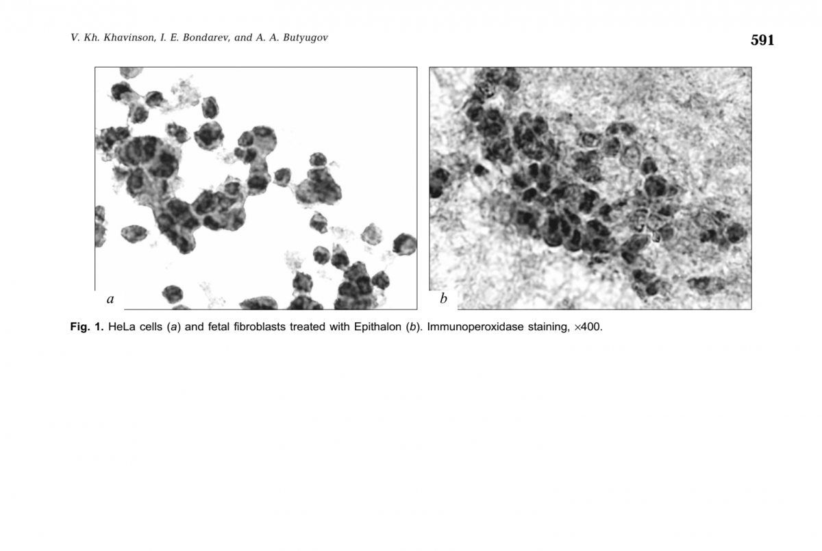
A long while ago I discussed this Blasco paper, where amongst other things they demonstrated that kidney cells with short telomeres undergo the EMT (epithelial to mesenchymal transition). This was reversed (MET) by extending telomeres. I speculated at the time of the importance for cancer, given the main source of cancer in humans is the epithelium.
I've also discussed before how with age cancer-like cells become more and more common, to the extend that I claimed 'cancer is aging.'


And as everyone knows by now, I'm a big fan of the telomere theory of aging - I think it's the lynchpin of all aging - and I've also never agreed that telomere shortening is all about senescent cells; like Michael Fossel I believe aging is analogue not digital, and that as telomeres get shorter cells become progressively more dysfunctional. It is not a sudden, all or nothing, distinction.


With that lengthy introduction out of the way…


Novel insights from a multiomics dissection of the Hayflick limit
https://elifescience.../articles/70283


This is a very important paper that recreated the original Hayflick experiments of passaging lung fibroblasts to replicative senescence, examining with a fine tooth comb changes in gene expression over time, and importantly controlling for cell density as well as contrasting wld type cells with the parallel changes in telomerase immortalised fibroblasts, as well as cells made senescent through radiation. As you read my highlights from the paper, you'll see what an important work it is, and why it's justified such a lengthy introduction.
I give you some choice quotes.


…the time resolution of our experiment coupled with single-cell trajectory analysis reveals that senescence is a gradual process that shares transcriptional, proteomic, and metabolomic features with epithelial-mesenchymal transition (EMT). Second, our metabolomic data identifies Nicotinamide N-methyltransferase (NNMT) activity as a potential initiating event in replicative senescence dependent loss of epigenetic silencing. Third, we show that these genomic regions that exhibit increased accessibility with increasing cellular age are concentrated in nucleolar/lamin associated domains and correspond with observed changes in the replicative senescence transcriptome...as expected, gene expression in the hTERT-immortalized WI-38 cells remained largely stable…the gene set for epithelial to mesenchymal transition (EMT) was significantly enriched in genes that increased with replicative senescence as opposed to radiation induced senescence…the EMT gene set is enriched early and robustly during replicative senescence…Interestingly, the vast majority of gene expression changes evident by PDL 50 begin to manifest at much earlier PDLs.


They carried out single-cell RNA experiments to see if the changes they were seeing was due to the changing mix of cells (some senescent, some not) or due to changes across all cells simultaneously. Remarkably they found the latter was true: the vast bulk of cells are moving together towards senescence. I cannot stress this last point enough. In their words.


Together, these results argue in favor of a gradual model of replicative senescence wherein cells ramp up expression of the replicative senescence program with increasing PDL even when still proliferative... Importantly, this observation suggests that aspects of cellular senescence are present in non-senescent cells. This result raises the intriguing possibility that the reported pathological features of senescent or senescent-like cells in vivo might also manifest in cells that are not classically senescent.'


From the proteomics data.


Multiple enriched sets pointed to replicative senescence-dependent shifts in cellular energy utilization…We observed PDL-dependent increase in proteins from all categories except for mitochondrial assembly and structures. These results are consistent with altered mitochondrial function…From these data, it is clear that WI-38 cells approaching replicative senescence undergo drastic shifts in metabolism. Specifically, we observed increased glucose utilization in glycolytic shunts coupled with an increase in fatty acid import and oxidation. It is possible that the senescent cells are switching to fatty acid oxidation to fuel increased TCA cycling and oxidative phosphorylation as glucose is diverted to macromolecule production….Likewise, consensus metabolomic findings in EMT models report increased TCA cycle products, altered lipid metabolism, and activated hexosamine pathway. These data provide functional metabolic evidence supporting a connection between replicative senescence, metabolic rewiring, and the increased expression of EMT-related genes.


The point about increased fatty acid oxidation is the opposite of that seen in the muscles of the elderly. This likely reflects difference consequences of aging in mitotic and post mitotic cells. But it matches what we know about cancer-primed cells increasing with age and shows this is due to telomere shortening. Moving on…


In review of our replicative senescence data for the metabolic and proteomic components of the NAD and methionine pathways, we found that one of the largest and earliest changes for any protein (or transcript) was the increased expression of Nicotinamide N-methyltransferase (NNMT)…DNA and histone methylation require abundant levels of the universal methyl donor S-adenosyl methionine (SAM). NNMT not only depletes SAM by catalyzing the removal of its methyl group, it does so by fusing the methyl group to the NAD+ precursor nicotinamide (NAM) resulting in the generation of methyl nicotinamide (MNA). Thus, NNMT effectively acts as a sink for two of the primary metabolites a cell requires for silencing chromatin and regulating gene expression…High NNMT expression has been implicated in SAM depletion in embryonic stem cells and cancer-associated fibroblasts (CAFs). In both cases, the functional consequences were similar; DNA hypomethylation and decreased capacity to form or maintain silenced loci and heterochromatin. The important point is that the loss of silencing is actively promoted through NNMT activity. One intriguing hypothesis is that increased NNMT activity with replicative senescence promotes loss of silencing and heterochromatin via SAM depletion.


In other words, NNMT upregulation depletes both NAD+ and SAM, affecting both acetylation and methylation of the genome. The chromatin areas that were most affected were associated with the lamina and nucleolus; these were the areas where the drop in SAM and NAD+ most increased chromatin accessibility. So what genes were most upregulated by these changes in the genome accessibility?


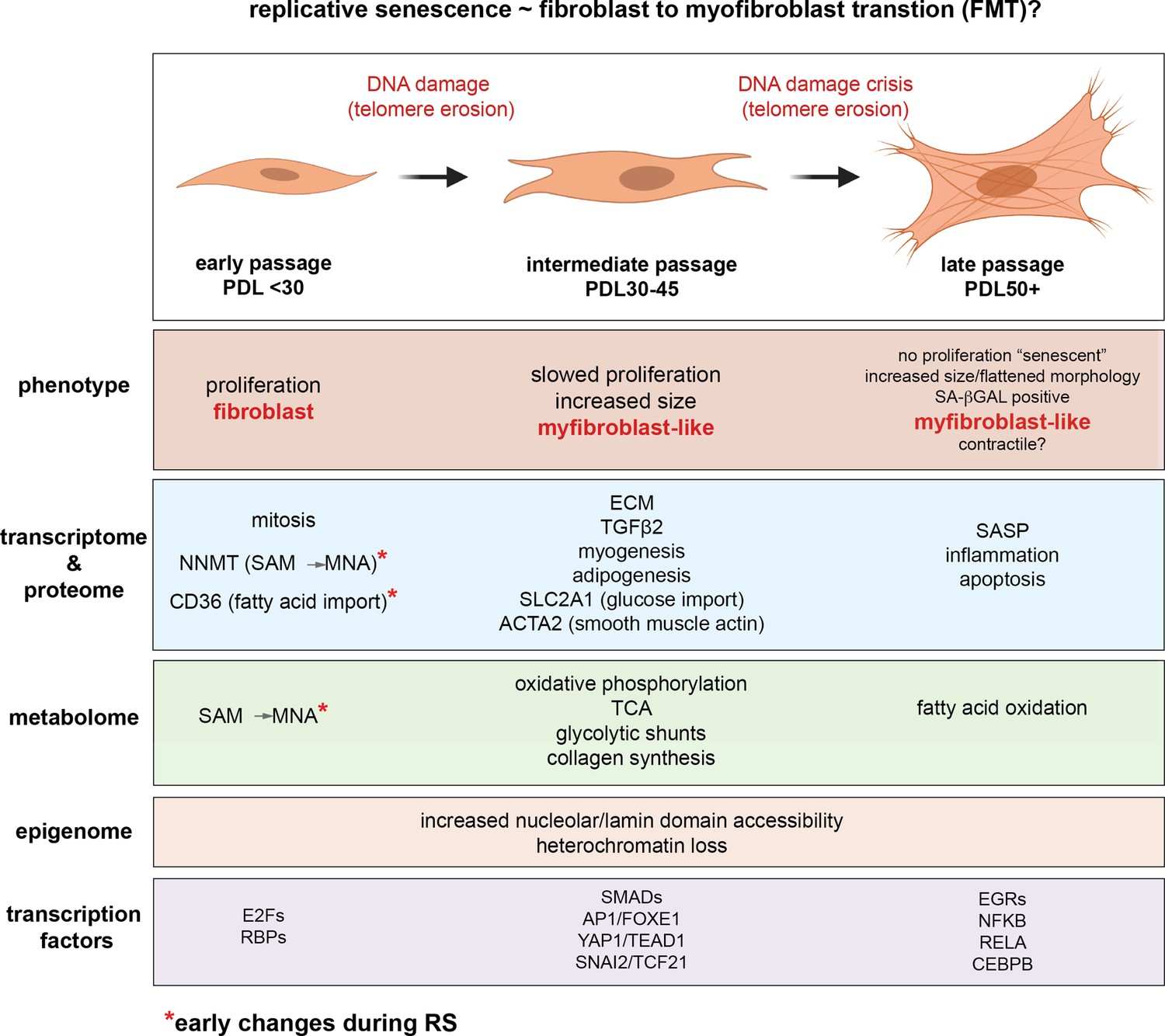
A large portion of the transcription factors exhibiting replicative senescence specificity belong to four categories: inflammation transcription factors (NFKB, CEBPB), AP1 subunits (JUN,JUND,FOSL2), YAP1-TEAD1 components (TEAD1/4, YAP1, WWTR1), and EMT transcription factors (SNAI2, TCF21)...'Collectively, these analyses uncover a common theme amongst putative regulatory transcription factors; Hippo signaling (YAP1/TEAD1), EMT transcription factors, and TGF-β signaling (SNAI2, SMAD activity). SNAI2 has been shown to work in tandem with the YAP1/TEAD1 complex and these pathways often work towards similar biological ends. Together, these transcription factors are reported as highly involved with proliferation, EMT, ECM production, fibrosis, and apoptosis avoidance…Collectively, these results present a possible order of operations for replicative senescence progression that highlights an initial cessation of active mitotic cycling, followed by an epigenetic shift that precedes a strong EMT/TGF-β signal before segueing into a pro-inflammatory secretory state…Given the repeated observations linking replicative senescence with EMT, TGF-β and YAP1/TEAD1 activity, we considered the possibility that these processes are connected through the fibroblast to myofibroblast transition (FMT), a subtype of EMT in which stressed or injured fibroblasts differentiate into myofibroblasts. Upon receiving cues mediated by injury or stress (e.g. activated TGF-β), fibroblasts can trans-differentiate into myofibroblasts, whose functions as ‘professional repair cells’ include increased proliferation, migration, apoptosis avoidance, cell and tissue contraction, and ECM/collagen deposition to promote tissue repair and wound closure…Previous work has demonstrated that there exists mechanistic and functional association between telomerase inhibition, senescence, and myofibroblasts. Senescence is an integral part of the wound healing processes; upon injury resolution, activation of a senescence-like phenotype prevents unchecked collagen secretion and fibrosis by preventing myofibroblast proliferation and earmarking them for subsequent immune clearance…Moving forward in our myofibroblast panel, we observed that expression of TGF-β cytokine, TGF-β1, decreased significantly with replicative senescence in both RNA and protein. However, we see robust induction of the TGF-β isotype 2 (TGF-β2) cytokine with replicative senescence as TGF-β1 abundance drops, which indicates a switch in TGF-β isotypes with replicative senescence. It has been shown TGF-β2 is a more potent inducer of the endothelial to mesenchymal transition (EndMT) in vitro compared to TGF-β1 and TGF-β3 in human microvascular endothelial cells, and TGF-β2 may be playing a similar role here in inducing FMT and replicative senescence in WI-38 cells.


It occurs to me that the beneficial action of TGF-B activating GDF11 may be converting transdifferentiated myofibroblasts back to the cell they should be, by rebalancing TGF-B isotypes. They also found a drug that blocks much of the TGF-B replicative senescence signalling through YAP1/TEAD1 (although I doubt this would be a plausible treatment for aging, see VERTEPORFIN).


Furthermore, we found that a subset of YAP1/TEAD1 targets are both induced with replicative senescence, and are principal components of TGF-β signaling. Thus, the YAP1/TEAD1 complex may be acting in convergence with TGF-β signaling with increasing PDL to enact a myofibroblast-like state that we recognize as replicative senescence…We found that treatment with 10 µM verteporfin for 2 hours reduced expression of these three YAP target genes by 70% relative to the no treatment control.


Some final choice quotes from the discussion section.


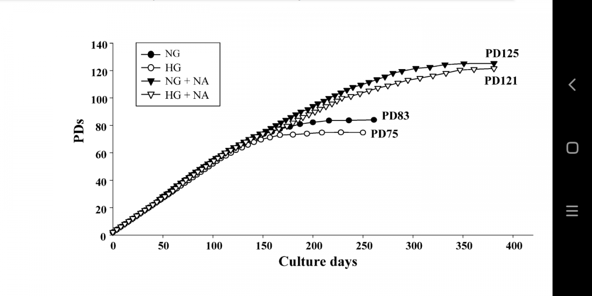
Importantly, very little changed across all modalities in the hTERT cell line.


No cellular aging if telomeres don't shorten.


…we provide evidence that the early manifestation of the senescent gene expression reflects gradual changes on a per cell basis rather than changing cell proportions. In effect, individual cells ‘show their age' with increasing PDL long before permanently exiting the cell cycle and transiting fully into the senescent state. The implications of this conclusion extend to organismal aging.


The importance of this paragraph for understanding aging cannot be overstated. In other words, it's no good just looking for specific markers for senescent cells. By inference senolytics will also not be a panacea.


Importantly, the gradual progression suggests that cells need not reach the endpoint to elicit a phenotype. For instance, proliferative fibroblasts isolated from IPF patients exhibited multiple senescent features and phenotypes in addition to accelerated senescence progression.


Perhaps measures of methylation aging are capturing some of this phenomenon, long before cellular senescence is reached.


Lastly, this phenomenon is not constrained to fibroblasts as we observe that the salient regulatory features of replicative senescence extend to cell types as distant as astrocytes.


It's not just fibroblasts. It is likely all cycling cells that suffer telomere loss.


We hypothesize that during fibrotic disease states and/or age, fibroblasts migrate to sites of micro-injuries. As these proliferating fibroblasts become replicatively aged, they are triggered (by DNA damage or other insults) to rewire their metabolism to induce FMT via active epigenetic reorganization.


In other words, aging is state whereby fibroblasts are trying to repair the body all over, leading to widespread conversion into myofibroblasts.


It is important to note here that the in vitro DNA damage here arises primarily from telomere erosion documented by the observation that our hTERT control cultures do not exhibit the same changes.


And there you have it. Despite barely mentioning telomeres until this point, this team have gone much of the way to proving the telomere therapy of aging. Thank you (Calico) and good night.


default.jpg


Edited by QuestforLife, 05 April 2022 - 03:55 PM.

  • WellResearched x 5
  • Informative x 2
  • Well Written x 1
  • Good Point x 1

sponsored ad

  • Advert

#754 QuestforLife

  • Topic Starter
  • Guest
  • 1,525 posts
  • 1,135
  • Location:UK
  • NO

Posted 07 April 2022 - 08:44 AM

The secret to lengthening telomeres in all cells: why I think epitalon doesn't activate telomerase and needs to be paired with a genuine activator


This information exists nowhere else; nowhere else on the internet and certainly in no peer reviewed journal. It may be the most important information I've shared on this thread. 


My definition of telomerase activation

Firstly, I need to be clear about what I mean by activating telomerase. There are various ways to measure telomerase. The most used is a TRAP assay, which measures the amount of detected telomerase protein. There is also a mRNA test to see how much telomerase RNA is being produced by the TERT gene. And of course we can measure telomeres at separate time points and measure the change in the telomere length distribution in those samples.

When I am talking about activating telomerase, I am talking about inducing gene expression to produce the mRNA, before it is made into the telomerase protein in the ribosome. The reason I stick to this definition is because at any given time you might have some telomerase protein but no new telomerase mRNA being transcribed from the gene. Or you can have telomerase RNA present but no manufacture (translation) into the finished protein by the ribosome factory. Or you might have telomerase RNA and protein present, but for some reason it is being diverted away from the telomere (say to the mitochondria in a time of high oxidative stress). That last possibility I will not discuss further as it can be addressed with anti-oxidants, inflammation control, etc.


Human Vs. in vitro Studies

It is often said that human studies are more important than anything else. After that are animal studies. And cell studies in a dish come a distant last.I generally agree, but studies at organism level often tell us nothing about mechanism and therefore can be misleading. For example a purported telomerase activator could extend life by a mechanism other than telomerase activation. Or a telomerase activator that has been shown to work in vitro to extend telomeres in cells might shorten life of the organism because of a separate effect it has on the body that did not affect the particular cell type tested in the dish. Obviously this argument extends to other substances operating through non telomere mediated mechanisms as well.


Why does any of this matter? Because I am concentrating on in vitro results where our range of variables is limited, and we can make reasonable deductions.


What do we know about epitalon?

Epitalon has been shown by studies in Russia to activate the telomerase (protein; note: not my definition of ‘activation’) and to extend telomeres [1], as well as extend the proliferative life of cells in culture [2]. This was done in human fibroblasts. Epitalon has also been shown to elongate the telomeres of leukocytes in many self experimenters (myself included), as well as in Russian studies [3]. Is this a slam dunk for peptide research and one in the eye for western researchers?


I am not so sure.


Bill Andrews has stated epitalon didn’t activate telomerase mRNA (my definition of ‘activation’) when he tried it (repeatedly). You could argue he is lying to sell more of his own telomerase activation product (TAM818). But that doesn’t make sense to me; he has been a telomerase researcher for many decades. If he heard of a substance that activated telomerase to the level of HELA cells (his Holy Grail), then he surely would be overjoyed if he could validate that claim regardless of his financials.


If we accept the above, epitalon must be acting at the cell level by increasing telomerase protein production from an already existing pool of telomerase RNA. Where would RNA this come from? My guess would be the calf serum used to maintain growing cells and re-added with each passage. This is fairly common practice, with greater or lesser control over contents. I ask you: is it likely small pieces of telomerase RNA could exist in the serum of unborn calves? I would be surprised if it wasn’t.  Something similar is done to conditionally immortalise somatic cells, namely the product of ‘feeder’ cells; these senescent murine ESCs don’t replicate but produce telomerase to ‘feed’ the non-telomerase producing human cells they share the culture with [3].


So much for the theory, where’s the evidence?

Is there any evidence for my theory that epitalon is using already present telomerase RNA to produce the protein? Maybe. If you look at Fig 1 from [1] (attached below), you’ll notice the HELA cells are dark with the detected telomerase protein against a white background (no reactivity). The epitalon-treated fibroblasts are also dark, showing the presence of telomerase protein within them, but their surroundings are also a mottled dark, showing the immunoperoxidase staining has also reacted with telomerase outside of the cells. They say (but do not show) that untreated fibroblasts do not have this staining (no telomerase present). Is it possible the mottled staining in Fig 1b is in fact telomerase RNA in culture? 


I’m not a bench scientist so have no way of doing a mRNA assay on fibroblasts treated with epitalon to verify no RNA is produced, and doing a parallel TRAP assay to show that telomerase protein was nevertheless produced, perhaps contingent on the inclusion of fetal calf serum. If you are and can do that work, please do so, you will deserve the plaudits that subsequently come your way.


Update! More Evidence!

I have been sitting on this post, thinking more and looking for more evidence.

I now think I have it. Peptides, including epitalon, are known to cause opening out of the chromosomes [4], [5]. It is called ‘decondensation’. It is interesting and relevant to this post because the regions that are decondensed allow expression of ribosomal genes. These are regions, conserved in most species, which produce the ribosomal RNA used to build the ribosome. This is the protein factory that makes all other proteins, including for our purposes, telomerase. This may explain the benefits of peptides in general: they are upregulating protein expression by building more ribosomes. In plain english, they turn on the factory that builds more factories. Whatever RNA is being produced by the cell in question, or hanging about in its cytoplasm, will be converted into the corresponding protein at a greater rate if stimulated by peptides. More ribosomal RNA equals more ribosomes and more protein expression.


The thread of my reasoning may seem tenuous. But think about this: what are peptides? Little pieces of broken up proteins. What would you expect to happen if lots of pieces of proteins got into a cell? The cell would increase protein assembly of course. That, I think, is what peptides do: they upregulate protein assembly. Now whether different peptides have different effects (as claimed) is an open question. Peptides are so small it is difficult to see epitalon (Al-Glu-Asp-Gly) being recognised as a piece of telomerase more than say another peptide with a slightly different amino acid makeup. Personally I think that is mostly sales spin. I can believe it for something super simple, like collagen. But telomerase is massive and really complicated (its structure still isn’t fully understood).


So that in a nut-shell is why I think epitalon will only work if the cells it is treating already have telomerase RNA present. In the case of leukocytes that is no problem, and explains why epitalon is so beneficial for the immune system (and lengthens telomeres according to the tests that only measure easily accessible immune cells). But really epitalon is only lengthening the telomeres of cells like leukocytes that (conditionally) make their own telomerase anyway - epitalon magnifies this.


Plan of Action

For self-experimenters who have access to telomere testing, there is something we could try.

Combining epitalon - hypothesized by me to increase (telomerase) protein assembly from RNA - with a proven telomerase ‘activator’ (mRNA activator that is), should produce a much greater increase in telomerase and increases in telomere length than either, used alone. Such a combination will have the ability to lengthen the telomeres of all cells, not just immune cells.


As far as I am aware TAM818, Product B (mix of nutraceuticals) and TA-65 have been shown to activate the HTERT gene to some level and should produce the required mRNA for epitalon to act on.


References

[1] https://pubmed.ncbi....ih.gov/12937682
[2] https://pubmed.ncbi....h.gov/15455129/
[3] https://pubmed.ncbi....h.gov/22189618/
[4] https://pubmed.ncbi....h.gov/16705247/
[5] https://pubmed.ncbi....h.gov/14647006/

Attached Thumbnails

  • epitalon effect on telomerase production in fibroblasts.png

Edited by QuestforLife, 07 April 2022 - 08:52 AM.

  • Informative x 3
  • Well Written x 1
  • like x 1

#755 QuestforLife

  • Topic Starter
  • Guest
  • 1,525 posts
  • 1,135
  • Location:UK
  • NO

Posted 07 April 2022 - 11:25 AM

I want to add a slight correction: the TRAP assay is a measure of the activity of the telomerase protein, i.e. if there is telomerase present it adds telomere repeats to a molecule that is then amplified and detected. The method used in Fig 1 attached to the post above was not a TRAP assay, it used immunostaining, whereby the ink sticks to the antigen selected, in this case the telomerase protein. This correction does not change the conclusions of post 754. 


  • Good Point x 1

#756 marcobjj

  • Guest
  • 310 posts
  • 26
  • Location:California

Posted 13 April 2022 - 03:42 AM

Do we know if the latest age reversal experiment done by Cambridge University is telomere related?


https://www.dailymai...s-30-years.html


#757 QuestforLife

  • Topic Starter
  • Guest
  • 1,525 posts
  • 1,135
  • Location:UK
  • NO

Posted 13 April 2022 - 08:10 AM

Do we know if the latest age reversal experiment done by Cambridge University is telomere related?

https://www.dailymai...s-30-years.html


Most of what academics do, even in such fields as epigenetic reprogramming, is irrelevant to life extension efforts. This is one such paper, where they took fibroblasts from middle aged people, gene edited them with a Doxycycline activator built in,and then used this to reprogram for longer than is normally done, i.e. the fibroblasts start to become pluripotent before they were allowed to revert to somatic cells. This meant a greater level of epigenetic reprogramming was acheived than is normally the case. But they still weren't reprogrammed for long enough for telomerase to be properly activated, as this only happens when cells are fully reprogrammed to pluripotency.

To investigate
the effect of transient reprogramming on telomere length, we used the telomere length
clock, which predicts telomere length based on the methylation levels at 140 CpG sites .
We found that MPTR does not affect telomere length and, in some cases, slightly reduces it
. This is consistent with our results profiling telomere length throughout
complete reprogramming using our doxycycline inducible system, where telomere length
did not increase until the stabilisation phase. This coincides
with the expression of telomerase during reprogramming, where it is weakly expressed
during the later stages of the maturation phase and only strongly expressed during the
stabilisation phase. Source:https://elifescience.../articles/71624


  • Good Point x 2
  • Informative x 1

#758 dlewis1453

  • Member
  • 151 posts
  • 45
  • Location:USA
  • NO

Posted 27 April 2022 - 03:33 PM

Are you familiar with the compound Tempol? Its an intracellular SOD mimetic that appears to have wide reaching and potent effects. The below site is a good repository for studies on Tempol. Tempol is a research chemical, not a supplement. It has been studied by researchers for a couple of decades now, but it is now in the spotlight again due to its antiviral properties. I thought it could help slow down telomere attrition through greatly improved regulation of oxidative stress. 


http://tempol.info/


Here is a description of Tempol: "Tempol is a redox cycling nitroxide that promotes the metabolism of many reactive oxygen species (ROS) and improves nitric oxide bioavailability. It has been studied extensively in animal models of oxidative stress. Tempol has been shown to preserve mitochondria against oxidative damage and improve tissue oxygenation."


Here is a study regarding Tempol and Telomeres, taken from the above site. Tempol does appear to benefit Telomeres. 


http://tempol.info/a...l-and-apocynin/


And another study that addresses Tempol and Telomeres. 


https://www.ingentac...000005/art00005


Here is a quote from Wikipedia:

"In biochemical research, 4-hydroxy-TEMPO has been investigated as an agent for limiting reactive oxygen species. It catalyzes the disproportionation of superoxide, facilitates hydrogen peroxide metabolism, and inhibits Fenton chemistry.[4] 4-Hydroxy-TEMPO, along with related nitroxides, are being studied for their potential antioxidant properties."


On the anecdotal front, one veteran user of Longecity "Hip" had his psoriasis permanently cured by Tempol. See his thread here: https://www.longecit...d-my-psoriasis/


  • Informative x 3

#759 QuestforLife

  • Topic Starter
  • Guest
  • 1,525 posts
  • 1,135
  • Location:UK
  • NO

Posted 28 April 2022 - 09:22 AM


Are you familiar with the compound Tempol? 


Thanks for posting this. I was not aware of this compound, although I have of course experimented with mitochondrial antioxidants like mitoQ. 


It is an open and very important question how much oxidative stress occurs to human cells In Vivo and how much this contributes to telomere shortening, either directly via damage to the telomeres, or by inducing cellular senescence via DNA damage elsewhere on the chromosomes, thereby necessitating their replacement by cell division, again causing telomere shortening. 


It is an unfortunate fact of life that the ROS produced by mitochondria cause telomere shortening above and beyond that caused by cellular division, and this also affects non or rarely dividing cells. It makes sense that a mitochondrial antioxidant could reduce or prevent this, provided this was done in a way that did not interfere with mitophagy, leading to a short term drop in ROS but a long term rise. I have spoken before about melatonin [1] being my top pick for a telomere-preserving mitochondrially-targeted antioxidant.


It is also of note that telomerase itself acts as an antioxidant, locating to the mitochondria in times of stress - which unfortunately stops it doing its job of lengthening telomeres in the nucleus.


Telomerase also increases mitophagy, so does not interfere with the mitochondrial maintenance programs that keep ROS from rising over time [2]. 


Bottom line: I am still unsure of the best approach to anti-oxidant use. It might even be that substances like nicotinamide that increase short term ROS, but delay its inevitable rise with age (caused by telomere shortening) are a better approach [3] - although they come with their own problems like reducing mitochondrial mass. 


[1] https://www.longecit...-16#entry905284

[2] https://www.longecit...-21#entry908174

[3] https://doi.org/10.1...26.2006.00234.x


Edited by QuestforLife, 28 April 2022 - 09:23 AM.

  • Good Point x 2
  • Informative x 1

#760 QuestforLife

  • Topic Starter
  • Guest
  • 1,525 posts
  • 1,135
  • Location:UK
  • NO

Posted 30 April 2022 - 03:11 PM

Tortoise vs. the Hare

If (T-cell) telomeres can have their telomeres lengthened by the equivalent of ~40 years loss in 4 years (and 40 years less that chronoAge; see attachments 1+2), but epigenetic age over the same period ends up at only 8 years less than chronoAge (attachment 3), what does that suggest?

It suggests to me that epithalamin peptide (and logically, epitalon) lengthens telomeres in both T cells and underlying stem cells. In the short term this leads to a dramatic increase in T cell telomere length. In the long term the more slowly dividing stem cells' telomeres are also extended. This then increases turnover and differentiation into T cells, which reduces T cells' epigenetic age. This is what you'd expect if T cells are being replaced more often because their progenitors are more able to divide, and is the opposite to what happens with age, i.e. shortening telomeres in stem cells slow the replacement of downstream cells (see attachment 4 of cell division rate falling with number of cell divisions).

So long term telomere extension should decrease epigenetic (methylation) age.

Depending on your age you may decide this is a wiser course of action than direct stem cells stimulation and expansion.

Interesting interviews of Dr.Bill Lawrence from where attachment s 1-3 are taken: https://youtu.be/NtLueX5qTBQ, https://youtu.be/3icPDEn6HpM

Attached Thumbnails

  • Screenshot_20220414-212208.png
  • Screenshot_20220414-212844.png
  • Screenshot_20220430-153523.png
  • Screenshot_20220430-145425.png

Edited by QuestforLife, 30 April 2022 - 03:17 PM.

  • Good Point x 1
  • Informative x 1
  • WellResearched x 1

#761 QuestforLife

  • Topic Starter
  • Guest
  • 1,525 posts
  • 1,135
  • Location:UK
  • NO

Posted 02 May 2022 - 08:55 AM

The secret to lengthening telomeres in all cells: why I think epitalon doesn't activate telomerase and needs to be paired with a genuine activator


Telomerase is made in the nucleolus

The nucleolus is formed in the nucleus when ribosomes (protein factories) need to be made. When ribosomes are complete they are sent out of the nucleus into the cytoplasm.

LRRC34 is a nucleolus protein also associated with longer telomere length both in humans [1]and with pluripotency and telomere length in stem cells[2],[3]

LRRC34 associates with nucleolin, another protein that exists in the nucleolus , which is known to retain telomerase in the nucleolus during its maturation [4]. It was news to me that telomerase was made in the nucleolus and not in normal protein manufacture once the ribosome has been made and sent into the cytoplasm. But it does make sense given the action of telomerase is primarily in the nucleus extending telomeres (notwithstanding it's mitochondrial actions in times of stress).

As I theorised previously, ribosomal upregulation increases the efficiency of mRNA translation, given the short half life of mRNA and the long half life of ribosomes. This paper shows it is not just building the (telomerase) protein by more ribosomes that increases this translation efficiency, but also has to do with how telomerase is sequestered and released at the right time for maximum effect.

Once again this shows how substances can increase telomerase protein and lengthen telomeres without activating the HTERT gene: by acting at a later stage.

It is still looking likely that epitalon can only increase telomerase in cells that already have a baseline production of the mRNA. Of course it may be that many human cell types do have small levels of telomerase mRNA production, say during S-phase, which is otherwise repressed. And this can be sequestered in the nucleolus. Nevertheless it is still my strategy to use another substance capable of activating the HTERT gene and use that in tandem with epitalon.

[1]https://doi.org/10.1...1386-018-0289-0
[2] https://doi.org/10.1089/scd.2021.0113
[3] https://www.liebertp...9/scd.2013.0470
[4] https://doi.org/10.1...tcb.2019.04.003
  • Good Point x 1
  • WellResearched x 1

#762 QuestforLife

  • Topic Starter
  • Guest
  • 1,525 posts
  • 1,135
  • Location:UK
  • NO

Posted 26 May 2022 - 03:09 PM

As a corollary to my post talking about telomerase RNA and telomerase protein detection (post#754), I thought I'd explain this a little more. There are actually 3 main methods for detecting telomerase, and I must admit that I was not fully clear on the distinctions. After reading this post you should be able to read a telomerase paper and understand what the authors mean.


PCR - polymerase chain reaction: should be familiar to anyone after 2 years of COVID. A primer (small single strand of DNA) is designed that will bind to its complementary target DNA sequence (once the DNA has split into 2 strands). Polymerases then add loose nucleotides from that point on to make the rest of the strand, so the DNA is doubled. Heat then splits the DNA again and once it's cooled enough the polymerases get back to work adding to where the primer has bound to. The whole process is repeated many times. This is called amplification. Eventually there is enough DNA to put it into an electrically charged gel that draws the DNA through it. Depending on the length of the DNA it is drawn through a greater or lesser amount and this is compared to a reference to see exactly how long it is and hence what it is.


For detecting RNA the process is basically the same only first the RNA has a primer and polymerase added to make it into DNA, then PCR happens as above.

So this is how you can detect telomerase RNA, i.e. that the telomerase gene has been turned on.


The TRAP (telomere repeat amplification protocol) is a way of detecting telomerase protein (once it has been converted or 'translated' from RNA in the ribosomes). The PCR and electrophoresis gel stages are identical to what I described above. The only difference is there is a very clever stage beforehand, whereby they add a length of nucleotides (single strand of DNA) to the solution (possibly) containing telomerase and if telomerase is present then it will recognise the 'oligonucleotide' strand and add telomere repeats to it. What is produced is then amplified by PCR and depending how much is produced then creates a mark that can be compared to a reference on the gel, which gives you an idea how much telomerase was present. 


In papers they should normally say they used TRAP, but sometimes it is merely referred to as telomerase 'activity' (i.e. the protein doing its job of lengthening (in this case fake) telomeres.


The third method is generally just referred to as measuring telomerase protein levels. I caution against relying on this method for reasons I will describe below.


This method is called immunohistochemistry. It relies on using an antibody that attaches to the protein in question (like immunity to a pathogen but in this case it is telomerase). The antigen will also be complexed with something else, often an enzyme. When the substrate is added to solution the enzyme causes a reaction to occur with its substrate, and this is what stains the protein. ELISA is a form of this.


Putting aside the RNA detection for a moment, a comparison has been made between TRAP and immunohistochemistry methods for detecting telomerase.


Comparison of NCL-hTERT Antibody Reactivity and Telomere Repeat Amplification Protocol in Situ in Effusions

Objective

To compare the performances of 2 methods, telomerase repeat amplification protocol (TRAP) in situ and antibodies to the hTERT protein, in assessing telomerase activity.

Study Design

TRAP in situ and immunohistochemistry with a commercial antibody (NCL-hTERT) was performed on 54 body cavity effusions. The results were compared and correlated to diagnosis.

Results

Thirty-four effusions from patients with verified malignant disease contained cytologically malignant cells. Both methods were positive in 33 of the cases, whereas only hTERT was positive in 1 case. Twenty effusions, all containing mesothelial cells, came from patients with benign conditions. In 2 fluids atypical, hyperplastic mesothelial cells were both TRAP in situ and hTERT positive. All remaining 18 fluids were TRAP in situ negative, whereas 12 of 18 were hTERT positive. Thus the results of TRAP in situ and hTERT immunohistochemistry disagreed in 1 of 34 (3%) malignant and 12 of 20 (60%) benign cases.

Conclusion
The sensitivities for malignancy were similar for TRAP in situ and hTERT immunohistochemistry. The specificity of the applied hTERT antibody was significantly lower, due to hTERT reactivity in mesothelial cells.
DOI: 10.1159/000325865


From the above we can see that sometimes benign tissue, which would be expected to be telomerase negative, can still come out positive according to immunohistochemistry, whereas only rarely do we get a false positive from TRAP.


My conclusion: HTERT protein levels as measured by immunohistochemistry are unreliable.


Are there any examples of this in the aging literature?


Resveratrol-induced augmentation of telomerase activity delays senescence of endothelial progenitor cells

DOI: 10.3760/cma.j.issn.0366-6999.2011.24.033


From the above document I attach the following images:

1. Telomerase mRNA; note 50uM resveratrol increases telomerase (mRNA) by approx. 70%

2. Telomerase protein as measured by immunohistochemistry; note 50uM resveratrol increases telomerase (protein) by approx. 140%

3. Telomerase activity as measured by TRAP; note 50uM resveratrol increases telomerase (activity) by approx. 78%


You can see immediately that number 2 - telomerase protein level as measured by immunohistochemistry - is out of step with the other two results and is in this case is overestimating telomerase production. 


Bottom Line


When looking at telomerase activation look for mRNA or TRAP and be suspicious of immunohistochemistry measurements of telomerase protein. 


Attached Thumbnails

  • Resveratrol_hTERT_mRNA.jpeg
  • Resveratrol_hTERT_protein_immunoBlotting.jpeg
  • Resveratrol_Telomerase_Activity_TRAP.jpeg

Edited by QuestforLife, 26 May 2022 - 03:18 PM.

  • Informative x 3

#763 QuestforLife

  • Topic Starter
  • Guest
  • 1,525 posts
  • 1,135
  • Location:UK
  • NO

Posted 14 June 2022 - 12:12 PM

Hyperfunctional Telomeres Part II:

How do you lengthen the telomeres of the small number of important stem cells, those cells responsible for the health of the whole organ in which they reside, in a way which will later restore lost youth to the body?


First a step back. I am operating from the assumption that the body is wholly dependent on replacement cells from stem cells, and that aging is basically stem cell telomere attrition. This matches with what we know of faster life strategies (and better early life performance) being correlated with an earlier onset of age related illness and death, association of amino acids with both faster aging and better early life performance, mTOR inhibition and CR being associated with longevity, etc.


Therefore, if stem cells are made to differentiate too often, this will shorten life, but self-renewal of stem cells will lengthen life.


It is important to note also that this is a dynamic process. It is not just a stem cell reserve being run down more or less quickly. If stem cells are given breathing space to renew, they can recover their telomeres, at least according to the worm data [1], and possibly for human cells too [2]. Therefore age reversal is possible.


I return to the earlier question: how do we get to those few, slowly dividing, almost quiescent stem cells on which the whole rest of the body depends?

We know the answer from the most successful anti-aging strategy: calorie restriction. Or the most successful pharmacological method for extending life: rapamycin (mTOR inhibition).


Both these methods reduce the differentiation of stem cells into tissues and upregulate self-renewal of stem cells. It has also been shown that saturated fats can bias cellular division towards symmetrical division, and that this can benefit stem cells residing in the gut [3], for example: in this sense fat burning may be a body wide signal for starvation. Antioxidants seem to have complicated effects on longevity, but even here we may understand their effects in terms of stem cell division, with antioxidants associated with improved performance (less age related mental decline) but shorter life [4]. 


A picture emerges that takes in all strands of gerontological research and is centered on stem cells and telomeres. It also suggests a potential strategy that has been espoused on this thread before [5], namely that we should be aiming to inhibit mTOR whilst boosting telomerase activity. This to me looks like the best bet for age reversal because it targets stem cell renewal whilst providing them with more of what they need to renew, namely telomerase. Recent increases in our understanding of the various stages of telomerase activation [6] also aid this cause.


Let me paint a picture of aging and how we might reverse it:

During life stem cells must choose between self-renewal and supplying the body with new cells. Generally evolution selects for faster cell replacement, and this deplete stem cell pools resulting in less cell replacement with age, hence aging.

mTOR inhibition or CR slows cellular division in the body, and allows stem cells to renew. During this time stem cells may actually be able to extend their own telomeres [1,2]. Adding additional telomerase at this time will increase this effect.


Later when normal growth signals resume, the longer telomeres of stem cells allow them to differentiate and renew the body at the rate they did when you were younger.


Experimental protocol to follow.


Ps: you can regard the hypothesis outlined in this post as an extension to Blagosklonny’s mTOR centric vision of aging. Pre-empting objections along the lines of ‘humans don’t generally die of aplastic anemia or pulmonary fibrosis therefore telomere shortening does not limit human lifespan’ - I would say you don’t die of these things because stem cells are sacrificing themselves so that you don’t. For example, analysis of an extremely old human revealed almost complete depletion of hematopoietic stem cells [7], even though she didn’t die of anemia.

[1] https://doi.org/10.1...mcr.2019.06.005
[2]  https://doi.org/10.3390/biom11030464
[3]  https://doi.org/10.1...tem.2018.04.018
[4] https://pubmed.ncbi....h.gov/22785389/
[5] https://www.longecit...-20#entry907024
[6] https://www.longecit...-26#entry915113
[7] doi:10.1101/gr.162131.113


Edited by QuestforLife, 14 June 2022 - 12:14 PM.

  • Informative x 3
  • WellResearched x 1
  • like x 1

#764 QuestforLife

  • Topic Starter
  • Guest
  • 1,525 posts
  • 1,135
  • Location:UK
  • NO

Posted 15 June 2022 - 09:13 AM

Summary of ‘Alternative Methods to Extend Telomeres’ Sept 2018 to Sept 2021 June 2022


Early work on NAD+
https://www.longecit...es/#entry857309
https://www.longecit...e-2#entry868202
SIRT4
https://www.longecit...e-3#entry870174
Loss of NAD+ because of telomere shortening
https://www.longecit...-13#entry900015


Work on Statin-Sartan protocol
https://www.longecit...es/#entry862269
link between ROCK inhibitors and telomerase
https://www.longecit...es/#entry864097
possible link with senolytics
https://www.longecit...es/#entry864534
using ROCK and mTOR inhibitors to reprogram brain cancer cells into normal neurons
https://www.longecit...es/#entry865160
How ROCK inhibitors block differentiation
https://www.longecit...e-4#entry878635
Feedback on protocol
https://www.longecit...e-5#entry881808
Summary of ROCK inhibition action on cells
https://www.longecit...e-6#entry883118
Attempts to come up with alternatives to statin and sartans
https://www.longecit...e-7#entry884915
Diagram of interventions
https://www.longecit...e-8#entry885663
Paper linking up ROCK and ECM
https://www.longecit...e-8#entry885731
ROCK and tgf-b
https://www.longecit...e-8#entry886244
Mean and Max lifespan extension with a ROCK inhibitor
https://www.longecit...-10#entry896988

Work on telomerase activators and other important telomere papers
Royal Jelly
https://www.longecit...e-2#entry866228
Review of various activators
https://www.longecit...e-4#entry875566
Asiaticoside
https://www.longecit...e-5#entry880274
Some other telomeres studies
https://www.longecit...e-7#entry884556
Effect of antioxidant on telomere shortening in the bone marrow
https://www.longecit...e-8#entry885539
More on the same, later
https://www.longecit...-10#entry896907
Telomere activators and CV diseases
https://www.longecit...e-8#entry885582
Telomere shortening predicts species life span
https://www.longecit...e-9#entry893160
using TERC upregulation to increase telomere length in stem cell
https://www.longecit...-10#entry896804
Telomerase and Splicing Factor regulators
https://www.longecit...-11#entry899109
T cells taking telomere length from other cells
https://www.longecit...-11#entry899161
Do stem cell stimulants deplete the bone marrow pool?
https://www.longecit...-13#entry900006
Hyperbaric oxygen therapy
https://www.longecit...-13#entry900378
Discussion of Blasco paper on hyperlong telomere mice
https://www.longecit...ndpost&p=901986
Discussion of actual in vivo rate of telomere attrition
https://www.longecit...-14#entry902137
GDF11 lengthens telomeres in MSCs via TERC upregulation
https://www.longecit...-15#entry903694
Possible benefit of Klotho to telomeres
https://www.longecit...-15#entry903694
Nucleotides (specifically guanine) for elongation of telomeres: eat Anchovies and Herring!
https://www.longecit...-15#entry904277
Blasco and short telomeres in kidney disease plus possible connection of short telomeres and the cancer causing epithelial to mesenchymal transition
https://www.longecit...-15#entry904567
What is the most powerful telomerase activator and a comparison of methods of measurement
https://www.longecit...-15#entry905188
Melatonin is the best antioxidant for telomeres?
https://www.longecit...-16#entry905284
More on melatonin
https://www.longecit...-18#entry906399
AKG and telomere length (in mice)
https://www.longecit...-16#entry905690
Discussion of a cell permeable, oxidation resistant form of Vit C and telomeres plus follow on discussion of ROS hormesis in some cell types
https://www.longecit...-16#entry905240
Various discussions on the bioavailability of Asiatic acid/asiaticoside (a purported telomerase activator) and why you may only want a very small dose
https://www.longecit...-14#entry903398
Should we be taking Zinc for our telomeres?
https://www.longecit...-17#entry906002

Clear benefits to life expectancy, CVD and Cancer with longer telomeres: a study with 500k people
https://www.longecit...-17#entry906042
Ability of endothelial cells to make new lining is telomere length dependent
https://www.longecit...-17#entry906088
Caffeine promotes telomerase expression
https://www.longecit...-17#entry906174
Dark chocolate for telomeres
https://www.longecit...-17#entry906249
Alternatives to a telomere test: NLR and CRP
https://www.longecit...-19#entry906563
Hyperfunctional telomerase: do you want more cell division or longer telomeres?
https://www.longecit...-20#entry907024
We should be aiming for mouse levels of telomerase, not HELA levels
https://www.longecit...-20#entry907165
New intranasal and injectable gene therapy for healthy life extension
https://www.longecit...-21#entry907730
New GDF11 telomerase paper in Nature:
Growth differentiation factor 11 attenuates cardiac ischemia reperfusion injury via enhancing mitochondrial biogenesis and telomerase activity
https://www.longecit...-21#entry907939
Telomerase increases mitophagy through PINK1 - explanations for my increased exercise tolerance
https://www.longecit...-21#entry908174
Polymorphic tandem DNA repeats activate the human telomerase reverse transcriptase gene
https://www.longecit...-22#entry908442
Telomere length and telomerase activity in T cells are biomarkers of high performing centenarians
https://www.longecit...-23#entry909206
Caffeine promotes the expression of telomerase reverse transcriptase to regulate cellular senescence and aging
https://www.longecit...-23#entry909654
Are the oncogenic effects of telomerase mediated by methyl transferases?
https://www.longecit...-23#entry910058
Loss of GDF11 shortens telomeres
https://www.longecit...-24#entry911007
Does Berberine shorten telomeres?
https://www.longecit...-24#entry911030
Mitochondrial Telomerase Reverse Transcriptase Protects from Myocardial Ischemia/reperfusion Injury 
https://www.longecit...-24#entry911292
Naringenin: super supplement?
https://www.longecit...-24#entry911334
NMN increases telomere length
https://www.longecit...-25#entry912192
Has the telomere theory of aging been proven?
https://www.longecit...-26#entry915078
Telomerase is made in the nucleolus
https://www.longecit...-26#entry915527
Telomerase protein levels as measured by immunohistochemistry are unreliable
https://www.longecit...-26#entry915527


View of Aging
Importance of cell size
https://www.longecit...e-4#entry877909
The Selfish Cell lives longer
https://www.longecit...e-5#entry880039
https://www.longecit...e-5#entry880339
Telomeres are NOT passive in aging
https://www.longecit...e-6#entry883065
Discussion of telomeres and cancer
https://www.longecit...e-9#entry892745
Senescence and Cancer, again
https://www.longecit...-10#entry897658
Are methylation changes with age evidence of a program?
https://www.longecit...-12#entry899778
Comments on heterochronic parabiosis
https://www.longecit...-13#entry900319
More on Selfish Cell theory of aging (2021)
https://www.longecit...-14#entry902349
Age related methylation and the connection with the Selfish Cell Theory of Aging
https://www.longecit...-16#entry905284
Plus why aging is cancer
https://www.longecit...-16#entry905627
Putting together telomere and hyperfunction theories of aging
https://www.longecit...-20#entry907460
Oxidative stress alters global histone modification and DNA methylation
https://www.longecit...-21#entry908138
How non-differentiating Selfish (stem) cells come to dominate the stem cell pool; links between methylation, telomerase and ROS
https://www.longecit...-21#entry908160
Finding the Culprit: the hormones required for sexual maturity may be the trigger that starts aging via downregulation of TET2
https://www.longecit...-22#entry908266
Discussion over whether methylation of gene promoters is protective against stem cell loss and the counter evidence: immortalised cells accumulate such methylation
https://www.longecit...-22#entry908322
Discussion of the combined use of telomerase activators, GDF11, AKG, vit A and C
https://www.longecit...-22#entry908398
Summing up the Twin Evils of aging
https://www.longecit...-22#entry908458
The Evolution of the Selfish Cell
https://www.longecit...-23#entry909231
Cancer and the Selfish Cell
https://www.longecit...-23#entry909457
Does the Selfish Cell imply programmed or accidental aging?
https://www.longecit...-23#entry909533
Finding the Culprit II: Species' cellular ROS level sets aging rate via down regulation of demethylases and failure of Circadian Rhythm
https://www.longecit...-23#entry909696
How does the Selfish Cell affect post-mitotic cells?
https://www.longecit...-23#entry909914
Why long telomeres won’t make you live forever, but short telomeres mean you’ll die young
https://www.longecit...-24#entry910244
Defining the steps that lead to cancer
https://www.longecit...-24#entry910636
What are epigenetic Aging tests actually measuring?
https://www.longecit...-25#entry912768
A new thesis
https://www.longecit...-25#entry914358
The Secret to lengthening telomeres in all cells
https://www.longecit...-26#entry915113
The Tortoise and the Hare
https://www.longecit...-26#entry915479
Hyperfunctional Telomeres Part II
https://www.longecit...-26#entry916386


Skin aging
Stem cell competition – can you have too much symmetrical division?
https://www.longecit...e-4#entry879560


Results
Methylation results from Statin-Sartan protocol
https://www.longecit...e-3#entry873678
Telomere length improvements via Lifelength
https://www.longecit...e-6#entry883063
PhenoAge improvements
https://www.longecit...e-6#entry883130
Epitalon increases methylation age and discussion
https://www.longecit...e-8#entry892505
Further discussion
https://www.longecit...-11#entry899397
https://www.longecit...-11#entry899496
https://www.longecit...-12#entry899538
Plan to reduce both telomere and methylation age
https://www.longecit...e-9#entry895170
No improvement in methylation age from 3 months of AKG
https://www.longecit...-10#entry896760
Improvement in methylation age from 6 months of AKG
https://www.longecit...-13#entry899822
Further improvement in epigenetic age (-6.6 years)
https://www.longecit...-14#entry903105
Summary of GDF11 experience with biomarkers
https://www.longecit...-15#entry905149
May 2021 Methylation age results
https://www.longecit...-20#entry907470
Discussion of reaction times on GDF11
https://www.longecit...-21#entry908189
October 2021 Trume Results
https://www.longecit...-24#entry910821
Amino Acid results: Do telomerase activators deplete glutamine?
https://www.longecit...-24#entry911858
February 2022 Trume Results
https://www.longecit...-25#entry914327


Sundry
Fatty Acid Oxidation
https://www.longecit...-10#entry896447
Starvation and stem cell renewal
https://www.longecit...-13#entry899839
See other thread:
Feeding stem cells: the strange case of dietary restriction and alpha lipoic acid
https://www.longecit...id/#entry885897
Possible use of pioglitazone with telomerase activators to increase subcutaneous fat without bladder cancer risk
https://www.longecit...-14#entry902929
Resveratrol is weird.
https://www.longecit...-16#entry905283
Demethylating the klotho promoter with hydrogen sulphide
https://www.longecit...-17#entry905925
Melatonin is linked to mitochondrial function and increases TET2 production
https://www.longecit...-21#entry908191
Discussion starting here on reversing thymic involution
https://www.longecit...-23#entry909243
Comment on gonadal rejuvenation by GDF11
https://www.longecit...-25#entry913693


  • WellResearched x 3
  • Informative x 1

#765 QuestforLife

  • Topic Starter
  • Guest
  • 1,525 posts
  • 1,135
  • Location:UK
  • NO

Posted 03 July 2022 - 08:43 AM

Telomere Shortening in Hematopoietic Stem Cell Transplantation: A Potential Mechanism for Late Graft Failure?

doi: 10.1053/bbmt.2002.v8.abbmt080597

A very moving paper covering two cases of patients who achieved successful implantation of HSCs into their bone marrow - required to replace their own - but later experienced failure of the production of peripheral blood cells.

Of these cases they show the first failure was likely due to the age of the donor. The ill child (7) had cancer and the treatments destroyed their marrow, and their grandmother (61) was the donor - whose telomeres whilst normal for their age, shortened rapidly in the host and were not equal to the demands of repopulation, hence failure two years after.

In the second case the donor was the ill teenager's sister. The recipient (13) had aplastic anemia, and the donor (14) it turned out had short telomeres for her age (possibly related to her brother's genetic condition). As a result, although everything went fine initially, 25 years later the recipient's blood cell counts began to fail.

In both cases you can see how repopulating the blood is highly demanding on the telomeres of the bone marrow and that normal life history also erodes them more gradually after this.

My feeling is that the bone marrow 'hides' the problem for the most part by keeping our blood counts up, and we do not realise the approaching shadow of death that travels from within the marrow outwards. Even in 'normal' aging devoid of the problems of these young patients, clonal expansion of just a few stem cell lines is ubiquitous after age 70 (https://doi.org/10.1...586-022-04786-y).

Does anyone still doubt how important telomeres are to aging?
  • Good Point x 1
  • Informative x 1

#766 johnhemming

  • Member
  • 311 posts
  • 122
  • Location:Birmingham, UK

Posted 03 July 2022 - 10:01 AM

Does anyone still doubt how important telomeres are to aging?


Telomeres are important, but you need to look at what encourages telomerase to function.


#767 QuestforLife

  • Topic Starter
  • Guest
  • 1,525 posts
  • 1,135
  • Location:UK
  • NO

Posted 03 July 2022 - 11:52 AM

Telomeres are important, but you need to look at what encourages telomerase to function.


Well this thread is devoted to such things. Without repeating huge amounts of information the pertinent check points are TERT mRNA expression, TERC mRNA expression,assembly of the complete telomerase protein in the nucleolus, trafficking of telomerase to the telomere rather than mitochondria, access of telomerase to the telomere itself (influenced by chromatin, the shelterin proteins and telomere length itself).

But I don't think you were asking a question, but rather having your own perspective to share?
  • Cheerful x 1

#768 johnhemming

  • Member
  • 311 posts
  • 122
  • Location:Birmingham, UK

Posted 03 July 2022 - 12:02 PM

But I don't think you were asking a question, but rather having your own perspective to share?


In the broadest sense the human telomerase reverse transcriptase gene (hTERT) which is the primary regulator is itself regulated by methylation and acetylation of the epigenome.  Hence if the epigenome is not in the right state for whatever reason hTERT will not be expressed.


Edited by johnhemming, 03 July 2022 - 12:02 PM.

  • Needs references x 1

#769 QuestforLife

  • Topic Starter
  • Guest
  • 1,525 posts
  • 1,135
  • Location:UK
  • NO

Posted 03 July 2022 - 12:10 PM

The flaws of methylation clocks

The flaws of methylation clocks are demonstrated by the following paper devoted to creating a methylation proxy for Telomere Length:

https://www.aging-us...cle/102173/text

Here they used various biobanks to measure TL and correlate it to CpG methylation at various genomic locations, combining them to form a methylation-telomere clock with an overall correlation with their TL measurements (themselves problematic) of 0.4-0.5 (not bad). The clock had better predictive power than their measurements of TL for various health outcomes (of the people those samples came from).

Problem with all this clever bio-informatics is they get more data but sadly no more understanding.

Later they looked at cell culture and found their clock advanced even in HTERT immortalised cells with no telomere shortening, showing they weren't actually measuring TL at all, but something else related to cellular replications. I've discussed what methylation-clocks are probably measuring before [https://www.longecit...5#entry912768].

The obvious flaw with these approaches is that if you reversed the age-related methylation of the telomere associated methylation clock you might think that you'd lengthened your telomeres. But based on this paper you'd probably be wrong.

Likewise just because, say, the Horvath methylation clock advances with age, doesn't mean that if you reverse it you reverse age, as methylation does not equal aging. Only with something mechanistically understood like telomeres can you infer cause and effect and predict the benefits of reversal of the aging trend in this biomarker.

But I seem to be talking to myself here as everyone else thinks methylation clocks actually are aging.
  • Good Point x 1

#770 QuestforLife

  • Topic Starter
  • Guest
  • 1,525 posts
  • 1,135
  • Location:UK
  • NO

Posted 03 July 2022 - 12:16 PM

In the broadest sense the human telomerase reverse transcriptase gene (hTERT) which is the primary regulator is itself regulated by methylation and acetylation of the epigenome. Hence if the epigenome is not in the right state for whatever reason hTERT will not be expressed.

Which is the case in almost all human cells, almost all the time, and what 'telomerase activators' attempt to address.

In terms of methylation, there is evidence that completely blocking SAMe with a competitor (sinefungin) can radically lengthen telomeres, but I'd doubt you'd survive having no methyl donors for long.

https://pubmed.ncbi....h.gov/34817067/

Edited by QuestforLife, 03 July 2022 - 12:17 PM.

  • Good Point x 1

#771 johnhemming

  • Member
  • 311 posts
  • 122
  • Location:Birmingham, UK

Posted 03 July 2022 - 12:19 PM

Problem with all this clever bio-informatics is they get more data but sadly no more understanding.


Actually  I agree with you.  I don't think changing the methylation clock does anything of any good.


My view is that aging of cells is specifically the senescence of the cells in the broader definition viz the "gradual deterioration of functional characteristics".  It occurs because either stem cells do not properly differentiate or somatic cells de differentiate.  This is constrained by a shortage of Acetyl-CoA in the nucleus of the cell which means the acetylation of the histone does not occur.


My own experimentation demonstrates that if you increase the availability of Acetyl-CoA then cells start functioning again.


Methylation patterns follow the acetylation process.


Looking at it simplisiically if a cell does not produce all the proteins it has evolved to produce it is not going to be as functional as it would be.


#772 Turnbuckle

  • Location:USA
  • NO

Posted 03 July 2022 - 12:32 PM


Looking at it simplisiically if a cell does not produce all the proteins it has evolved to produce it is not going to be as functional as it would be.


You don't want cells producing all the proteins they are programmed for genetically. That would be disastrous. You want them producing only the proteins directed by the epigenetic code. And neither Acetyl-CoA nor any other supplement can fix it once it's mutated. Telomeres and the suicide genes activated by short telomeres are an evolutionary workaround. Since the body cannot detect or fix epigenetic errors, it slaps a Hayflick sell-by date on cells in the form of telomeres.


Edited by Turnbuckle, 03 July 2022 - 12:35 PM.


#773 QuestforLife

  • Topic Starter
  • Guest
  • 1,525 posts
  • 1,135
  • Location:UK
  • NO

Posted 03 July 2022 - 01:03 PM

You don't want cells producing all the proteins they are programmed for genetically. That would be disastrous. You want them producing only the proteins directed by the epigenetic code. And neither Acetyl-CoA nor any other supplement can fix it once it's mutated. Telomeres and the suicide genes activated by short telomeres are an evolutionary workaround. Since the body cannot detect or fix epigenetic errors, it slaps a Hayflick sell-by date on cells in the form of telomeres.

My own view of methylation-clocks is that they are a measure of how well the cell's methylation machinery is keeping up after DNA replication or repair. Depending on what you did you could fix this without reversing aging. That was the basis of my criticism of the current 'worship' of methylation-clocks above.

The fact is though, that HTERT immortalisation keeps somatic cell gene expression stable regardless of the advance of methylation-clocks in those cells (https://www.longecit...-26#entry915078) So it's going to be a hard task to argue causation from the methylation based changes measured by those clocks to aging.

Of course you could argue (and you do) that methylation-clocks are just a measure of (the failure of) stem cell replacement,and that is a much more satisfactory mechanistic explanation as those downstream cells could have all sorts of accumulated problems.

Edited by QuestforLife, 03 July 2022 - 01:05 PM.


#774 johnhemming

  • Member
  • 311 posts
  • 122
  • Location:Birmingham, UK

Posted 03 July 2022 - 01:29 PM

Whatever the mechanism of aging is, it has to explain all the known facts.


We know that heterochronic parabiosis has an effect (as does plasma replacement).  Methylation benig at the core of aging does not explain that.  Hence Methylation is not at the core of aging.


  • Needs references x 1

#775 johnhemming

  • Member
  • 311 posts
  • 122
  • Location:Birmingham, UK

Posted 03 July 2022 - 01:33 PM

You don't want cells producing all the proteins they are programmed for genetically. That would be disastrous. You want them producing only the proteins directed by the epigenetic code. 


I perhaps was not clear.  When I say "it has evolved to produce" I mean assuming the cell has differentiated.  The problem is when the cell does not differentiate properly and perhaps only produces some of the proteins.  


#776 Turnbuckle

  • Location:USA
  • NO

Posted 03 July 2022 - 01:57 PM

Of course you could argue (and you do) that methylation-clocks are just a measure of (the failure of) stem cell replacement,and that is a much more satisfactory mechanistic explanation as those downstream cells could have all sorts of accumulated problems.


I do. And because the cells has no means of making an epigenetic clock, it uses the simple telomeric clock. 


Whatever the mechanism of aging is, it has to explain all the known facts.


We know that heterochronic parabiosis has an effect (as does plasma replacement).  Methylation benig at the core of aging does not explain that.  Hence Methylation is not at the core of aging.


It appears that young blood contains a stem cell stimulant and/or rejuvenat --


The young circulatory milieu capable of delaying aging in individual tissues is of interest as rejuvenation strategies, but how it achieves cellular- and systemic-level effects has remained unclear. Here, we constructed a single-cell transcriptomic atlas across aged tissues/organs and their rejuvenation in heterochronic parabiosis (HP), a classical model to study systemic aging. In general, HP rejuvenated adult stem cells and their niches across tissues. In particular, we identified hematopoietic stem and progenitor cells (HSPCs) as one of the most responsive cell types to young blood exposure, from which a continuum of cell state changes across the hematopoietic and immune system emanated, through the restoration of a youthful transcriptional regulatory program and cytokine-mediated cell-cell communications in HSPCs. 

https://www.cell.com...(22)00170-9.pdf


If you stimulate stem cells and they replace old cells more rapidly, you get a reversal of epigenetic age, as cells derived from SCs have a low epigenetic age and drive down the average. The result lasts a few months in these rodents. So if this parabiosis experiment were performed repeatedly to improve the epigenetic age reversal, would stem cell niches get depleted? This experiment has not been performed to my knowledge, but I think it would. Stem cell depletion explains the failure of the Moody group to reproduce the 2012 results of the Moussa group. There was a hidden variable unappreciated by both groups, and that was mitochondrial fusion. Mito fusion is easily achieved by fasting in rodents, which have a metabolic rate six times higher than humans. The Moussa group likely fasted their rodents overnight (as they did in their toxicity study) but failed to mention it in regard to their longevity study, thus the Moody group didn't do it and the experiment failed. (I asked them about it by email, but didn't receive a response.)


Edited by Turnbuckle, 03 July 2022 - 02:13 PM.


#777 QuestforLife

  • Topic Starter
  • Guest
  • 1,525 posts
  • 1,135
  • Location:UK
  • NO

Posted 03 July 2022 - 02:45 PM

I do. And because the cells has no means of making an epigenetic clock, it uses the simple telomeric clock.


It seems strange that methylation age is such a good clock, but cells themselves can't use it. Seems contradictory to me. Instead I think cells probably do use it. Methylation might be one of the ways stem cells control differentiation (in addition to the mitochondrial controls you discuss).

It appears that young blood contains a stem cell stimulant and/or rejuvenat --


If you stimulate stem cells and they replace old cells more rapidly, you get a reversal of epigenetic age, as cells derived from SCs have a low epigenetic age and drive down the average. The result lasts a few months in these rodents. So if this parabiosis experiment were performed repeatedly to improve the epigenetic age reversal, would stem cell niches get depleted? This experiment has not been performed to my knowledge, but I think it would. Stem cell depletion explains the failure of the Moody group to reproduce the 2012 results of the Moussa group. There was a hidden variable unappreciated by both groups, and that was mitochondrial fusion. Mito fusion is easily achieved by fasting in rodents, which have a metabolic rate six times higher than humans. The Moussa group likely fasted their rodents overnight (as they did in their toxicity study) but failed to mention it in regard to their longevity study, thus the Moody group didn't do it and the experiment failed. (I asked them about it by email, but didn't receive a response.)


Problem is symmetrical division increases stem cell numbers but decreases their telomere length. Therefore your efforts may be falling into the same trap by increasing healthspan but not lifespan. I hope not, but there we have it.
  • Good Point x 1

#778 johnhemming

  • Member
  • 311 posts
  • 122
  • Location:Birmingham, UK

Posted 03 July 2022 - 04:11 PM

It appears that young blood contains a stem cell stimulant and/or rejuvenat --


a) I disagree

b) How would this affect methylation?


#779 Turnbuckle

  • Location:USA
  • NO

Posted 03 July 2022 - 04:19 PM

It seems strange that methylation age is such a good clock, but cells themselves can't use it. Seems contradictory to me. Instead I think cells probably do use it. Methylation might be one of the ways stem cells control differentiation (in addition to the mitochondrial controls you discuss).


The clock was created by researchers. It doesn't exist in the cell. And stem cells have very little methylation.


Edited by Turnbuckle, 03 July 2022 - 04:21 PM.

  • Good Point x 2

#780 QuestforLife

  • Topic Starter
  • Guest
  • 1,525 posts
  • 1,135
  • Location:UK
  • NO

Posted 21 July 2022 - 09:40 AM

What is Tianshengyuan-1?


Primary references:

1. Effect of Tianshengyuan-1 (TSY-1) on telomerase activity and hematopoietic recovery - in vitro, ex vivo, and in vivo studies

https://www.ncbi.nlm...les/PMC3992398/

2. Tianshengyuan-1 (TSY-1) regulates cellular Telomerase activity by methylation of TERT promoter

https://www.ncbi.nlm...les/PMC5352375/

3. Cellular senescence and cancer: Focusing on traditional Chinese medicine and natural products

https://doi.org/10.1111/cpr.12894

4. Schisandrin A and B affect the proliferation and differentiation of neural stem cells

https://www.scienced...891061821001411


This liquid herbal extract has been shown to activate telomerase in CD34+ hematopoietic stem cells but inhibit it in HL60 cancer cells. It is used in China for aplastic anemia, so it seems to be effective in vivo.


I attach some pictures of the effects it has on telomere length and telomerase activity. Apologies for the crude cut and paste job. All pictures are taken from references 1-4.


If Tianshengyuan’s specific telomerase boosting effects in stem cells could be confirmed, it would make a valuable addition to our arsenal against aging. Trouble is I’ve never even seen it for sale in the West, and it is not clear what it is composed of. According to reference 3 (Cellular senescence and cancer: Focusing on traditional Chinese medicine and natural products):


Tianshengyuan-1 (TSY-1) is exacted via a distillation process of multiple Chinese herbs, involving magnolia officinalis, schisandra Chinensis, pericarpium citri reticulatae viride and almond.


Of these schisandra is known for a positive effect on telomerase, see reference 4, with the effects not being as strong as claimed for TSY-1, at least in the neural stem cells used here, and with a much lower dose.This supplement at least is available more widely, although it is not always clear what percentage is made up of the active compounds Schisandrin A and B. 


If anyone has knowledge of Tianshengyuan, please share it here.

Attached Thumbnails

  • TSY effect on HL-60, PBMCs and HSC.png
  • TSY effect on HSCs.png
  • TSY effect on HSCs in a mouse model of aplastic anemia.png
  • TSY effect on T cells taken from Humans.png
  • TSY effect on HSCs taken from humans.png
  • Schisandrin A and B effect on telomerase in neural stem cells at 0.ug per ml.png

Edited by QuestforLife, 21 July 2022 - 09:46 AM.

  • Informative x 2


Also tagged with one or more of these keywords: telomeres, nad, nampt, ampk, resveratrol, allicin, methylene blue, nmn, sirtuins, statin

0 user(s) are reading this topic

0 members, 0 guests, 0 anonymous users